The Rise of Theranostics: A 40-Year Journey
January 15, 2025
Renowned theranostics expert discusses the evolution of the field and its expanding applications.

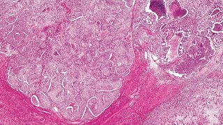
The Division of Nuclear Medicine, working with the UT Southwestern Harold C. Simmons Comprehensive Cancer Center, has made significant inroads into molecular imaging and therapy for cancer.
Martin Pomper, M.D., Ph.D., Professor and Chair of Radiology at UT Southwestern, has dedicated over three decades of his life to the development and translation of molecular imaging and theranostic (e.g., therapeutic and diagnostic) agents. His work led to the U.S. Food and Drug Administration’s approval of the first commercial prostate specific membrane antigen (PSMA) imaging agent, PYLARIFY® (piflufolastat F 18), in 2021. A drug with a similar chemical structure, PLUVICTO® (lutetium Lu 177 vipivotide tetraxetan), which is a PSMA-targeted molecular radiotherapeutic, was approved in 2022. Both are used widely for diagnosing and treating prostate cancers, respectively.
The field of theranostics is not new. It goes back to the 1940s with the treatment of thyroid cancer with radioactive iodine. By the 1980s, receptor-based imaging and therapeutic agents – for example, those targeting the estrogen receptor (ER) to image and potentially treat ER-positive breast cancer –were under development. My initial graduate thesis project was to develop Auger electron emitters targeting the ER.
Martin Pomper, M.D., Ph.D.
Discovering the Target
As a junior faculty member at Johns Hopkins, Dr. Pomper was interested in developing positron emission tomography (PET) imaging agents to study glutamatergic neurotransmission in the brain.
“Back in those days I would find targets for imaging by reading the Journal of Medicinal Chemistry or Cancer Research,” Dr. Pomper says. “One that caught my eye was about high-affinity inhibitors for a brain enzyme that generates glutamate, called N-acetylated α-linked acidic dipeptidase (NAALADase).”
Upon further investigation, Dr. Pomper discovered that the brain enzyme was homologous to PSMA. From there, he and his team pivoted from focusing on NAALADase in the brain to PSMA in the periphery, specifically to target prostate cancer. Over the next two decades, his group developed a variety of radiohalogenated and radiometalled agents to image and treat prostate cancer by targeting PSMA.
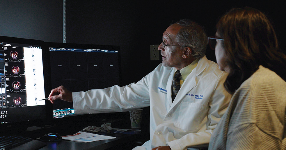
“We discovered that the radioactive small molecules we developed could image PSMA with high sensitivity and specificity,” Dr. Pomper explains. “Conventional bone and CT scans are being replaced by PSMA PET scans. The primary indications for PSMA PET are biochemical recurrence, namely, when a patient who has had a prostatectomy presents with a rising prostate specific antigen (PSA) level and there is a need to rule out a systemic recurrence, and for staging patients with high-risk disease.”

Commercial Interest Takes Off
According to Dr. Pomper, the acquisition of two startups by a large pharmaceutical company was a key milestone in the renaissance of molecular imaging and therapy for cancer, ultimately leading to the FDA approval of LUTATHERA®, for neuroendocrine tumors, in 2018 and PLUVICTO® in 2022.
“Because of that recent commercial interest, the field has skyrocketed in growth,” he says, with dozens of new companies dedicated to theranostics forming continually. “As for imaging, PYLARIFY® remains a dominant force in the U.S. market. There is also a growing market in Australia, New Zealand, and, more recently, in Europe. Other imaging and theranostic agents targeting PSMA – as well as a wide variety of other key targets in cancer – are under development worldwide.”
Collaboration Will Foster Innovation
Beyond PSMA-targeted radionuclide therapy, Dr. Pomper and his team are also looking at a number of imaging agents to evaluate neuroinflammation and biological processes in the central nervous system. Moreover, he has a keen interest in applying artificial intelligence in radiology, with a focus on using precision imaging agents to predict outcomes and select patients for appropriate therapies.
“In addition to the proliferation of precision imaging and theranostic approaches, in the future, minimally invasive procedures will also become increasingly common so that the work of interventional radiologists will begin to converge with that performed today by surgeons,” Dr. Pomper says. “Precision image-guided, robot-assisted biopsies will be routine. We will see more quantitative lesion characterization through photon-counting CT, MR fingerprinting, and an array of new molecular imaging agents against targets identified through proteogenomics.”
Dr. Pomper explains that that these advances will result from transdisciplinary collaboration among fields, including biomedical engineering, materials science, chemistry, computer science, informatics, and medicine.
Martin Pomper, M.D., Ph.D., is Chair and Professor of Radiology at UT Southwestern. He holds the Effie and Wofford Cain Distinguished Chair in Diagnostic Imaging. He is an organic chemist specializing in the development and clinical translation of radiopharmaceuticals and molecular radiotherapeutics. He is also a Professor in the Advanced Imaging Research Center and the Department of Biomedical Engineering and a member of the Chemistry and Cancer Research Program at the Simmons Cancer Center.
Dr. Pomper is a coinventor on a U.S. patent covering PYLARIFY® and, as such, is entitled to a portion of licensing fees and royalties generated by this technology. This arrangement has been disclosed to UT Southwestern in accordance with its conflict-of-interest policies.