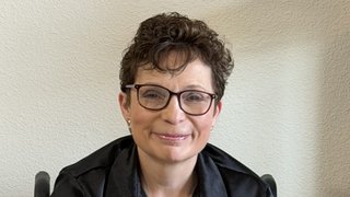
Dr. Elizabeth Maher, M.D., Ph.D.
“This is a solvable problem. You just need the right mix of people and a little bit of good luck.”
Professor of Neurology and Internal Medicine
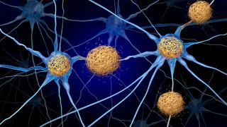
Dr. Elizabeth Maher spends much of her time thinking about the brain. To her, the brain is like a black box; the answers are in there, but getting to them and deciphering their precious codes are monumental tasks. It’s this type of thinking that ultimately spurred her initial engagements with imaging scientists and radiologists at UT Southwestern and led to groundbreaking advancements in neuro-oncology research.
Her attraction to the field of neuro-oncology stemmed from a personal longing to enrich the lives of patients facing terminal illness. The way she viewed it, not only was neuro-oncology a perfect fit for her clinical aspirations, but also a field in which “any little increment could make a gigantic difference.” As fate would have it, she found herself surrounded by a mix of people with equally inquisitive minds who helped produce work that would indeed make quite an impact.
“The key to our entire program is that Dr. Malloy knew what people were doing and could see there was a potential for collaboration that might be useful.”
Dr. Elizabeth Maher
The Collaboration
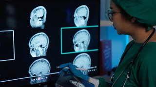
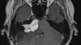
Dr. Maher’s novel idea, which was spurred by Dr. Craig Malloy (Medical Director of the Advanced Imaging Research Center), was to collaborate with scientists in UT Southwestern’s Radiology Department to see if a common gene mutation (IDH1) found in patients with glioblastoma tumors could be identified via advanced MRI scans – as opposed to the traditional method of extracting brain tissue via high-risk surgeries.
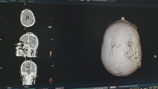
The collaboration was a complete success, thanks in large part to the expertise of the imaging team, led by Dr. Changho Choi, and the department's highly sophisticated 7 Tesla MRI. The team was able to identify the presence of the IDH1 mutation without the need for invasive surgery, and for the first time in medical history, a biomarker had been identified solely with MRI technology.
Imaging
Dr. Choi is a physicist in UT Southwestern’s Advanced Imaging Research Center whose work with magnetic resonance spectroscopy has helped improve the lives of neuro-oncology patients. He continues to collaborate with Dr. Maher and other neuro-oncologists as they work toward developing a fast, high-precision 2HG imaging modality.
“You take a group of patients who had no option – zero, don’t even know what to treat them with – and make them eligible for clinical trials. That’s a huge leap.”
Dr. Elizabeth Maher
The Importance of IDH
IDH is a mutation found in glioblastoma tumors that comes in two forms, IDH1 and IDH2. The mutation helps doctors determine the state of a tumor and is a key target for trial drugs aimed at inhibiting its influence on tumor growth. Glioblastomas that contain the IDH mutation start out as low-grade and slow–growing. Traditional methods of identifying IDH’s presence in brain tumors involved extracting brain tissue via surgery, which is both a lengthy and often dangerous procedure.
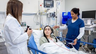
The collaboration between Dr. Maher and Dr. Choi revolutionized this process by identifying via advanced magnetic resonance spectroscopy the chemical signature of a metabolite that increases due to the presence of an IDH mutation, called 2HG. If 2HG is detected in a tumor, then it follows that the patient has the IDH mutation. If 2HG is not detected, doctors can conclude the patient does not have an IDH mutation. Because of the precision of the method, patients with 2HG detected by imaging can now immediately be placed into clinical trials using IDH inhibitors without undergoing invasive surgery. This is especially beneficial for patients with tumors located in high-risk regions of the brain.

“Even [for] patients with an excellent prognosis... these are still incurable, and we have to make that final breakthrough discovery that’s going to cure these tumors before they progress.”
Dr. Elizabeth Maher

The Impact
If it weren’t for Dr. Maher’s collaboration with Dr. Choi and his team of physicists and radiologists, many glioblastoma patients would be left in a constant state of wonder about the severity of their tumor and would not be able to receive proper treatment. Thanks to the advancements made with sophisticated MRI technology, patients battling brain tumors are now finding better treatment options faster.
It’s an impressive feat that’s improved the lives of many suffering from the disease, but Dr. Maher and her team know that this is just one step on the journey toward complete eradication. More importantly, they know that a key to reaching this goal lies in the collaboration between passionate individuals from a vast array of fields. As they continue adding individuals to the mix, they hope you consider joining them on this journey toward a cure.