Patience is key: Understanding the timing of early ultrasounds
April 8, 2025

The excitement newly pregnant women have to see how their baby is doing via an ultrasound can send them to an Ob/Gyn quickly. However, it’s important that they’re patient when their doctor recommends waiting until they are six weeks pregnant for their first ultrasound. Here’s why.
The First Visit to the Ob/Gyn
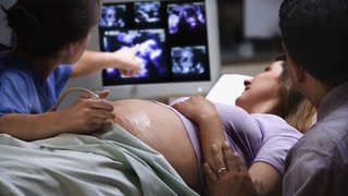
The first step women usually take after learning they’re pregnant is visiting with an Ob/Gyn. During this visit, an ultrasound is frequently done to confirm early pregnancy.
What to Expect from an Early Ultrasound
An ultrasound doesn’t immediately show what women might expect. It’s typically not until a woman is six weeks pregnant that any part of the fetus is visible, which allows the doctor to determine whether a pregnancy will be viable.
Because of this, it’s important that women understand what information their ultrasound can and cannot provide at certain times during their pregnancy. And they should be prepared to wait to learn more about their baby, if it’s too soon.

4 stages of early pregnancy and what we might see on ultrasound
An ultrasound is a routine part of prenatal care at six to nine weeks. The ultrasounds we might do prior to that, and the information those exams would reveal, generally occur in four stages:
- Stage One: If performed around the time a women’s menstrual period is expected, this ultrasound typically shows a fluffy, thick lining of the uterus that’s preparing for the fertilized egg to implant.
- Stage Two: This is usually at four to five weeks after a pregnant woman’s last period. The ultrasound commonly shows a small collection of fluid within the lining of the uterus that represents the early development of the gestational sac.
- Stage Three: This is usually about five and a half weeks after a pregnant woman’s last period. The ultrasound typically shows a gestational sac and within it we can see a 3-5 mm bubble-like structure, which is the yolk sac.
- Stage Four: Approximately six weeks after a pregnant woman’s last period, we can see a small fetal pole, one of the first stages of growth for an embryo, which develops alongside the yolk sac.
While these are the expected times to see the developing pregnancy with an ultrasound, not all pregnancies develop along the same timeline. Just like pregnancy tests, if there’s variability in the length of the menstrual cycle or when fertilization takes place, then what we see on ultrasound can change. It’s important that this ultrasound is performed vaginally for high-quality pictures.
If the first ultrasound doesn’t show a developing baby with a heartbeat, when should the next one be scheduled?
Newly pregnant women get anxious if we don’t see both a fetus and a heartbeat on the first ultrasound and frequently want to come back soon after for another look. But it takes time to move through the early stages of pregnancy. And repeating an ultrasound still won’t be able to reassure the patient that the fetus is alive and growing, if we do it too soon.
The general recommendations are to wait two weeks if we only see a gestational sac and at least 11 days if a gestational and yolk sac are seen without a fetal pole. I prefer to wait two weeks for the next ultrasound in both of these scenarios. If we can see an early fetal pole but can’t see cardiac movement, then we repeat an ultrasound in one week. I know waiting is hard – but in my experience, it is much better to wait and get a definitive report on the status of your pregnancy than potentially have to come back multiple times.
Pregnancy Loss
Pregnancy loss can occur during any stage of pregnancy, though it’s most common in the first trimester. If we do an ultrasound and the length of the baby is more than 7mm, we should always see movement of the fetal heart. If we don’t, we know the pregnancy is not going to develop.
"It’s important that women understand the right time to visit their Ob/Gyn for an ultrasound, so they can know with more certainty what to expect during the exam."
– Robyn Horsager-Boehrer, M.D.

Sign up for DFWChild’s Week-by-Week Guide to Pregnancy, presented in partnership with UT Southwestern Medical Center, to get timely information and updates.
When might earlier ultrasounds be recommended?
If you have a history of tubal pregnancy, the most common being an ectopic pregnancy, or when the fertilized egg implants outside the uterus, or develop symptoms that are concerning for the possibility of a tubal pregnancy, your doctor might perform an ultrasound sooner. In this instance, the doctor is looking for any evidence of a pregnancy within the uterus, so a gestational sac with a yolk sac is reassuring and means that the chances of a tubal pregnancy are low. They also will likely want to take a close look at the fallopian tubes to see if there is any sign of a mass in the tube that might represent a growing tubal pregnancy.
Although it is hard to be patient, understanding the early timeline for a developing pregnancy will help you understand when first trimester ultrasounds should be performed.
Related reading: Helping you understand scary (but often harmless) pregnancy ultrasound findings