Combining attentive, compassionate care with our extensive clinical and research resources, UT Southwestern's cardiology experts and vascular specialists deliver individualized care within pre-eminent healthcare facilities.
Noninvasive Vascular Imaging
New Patient Appointment or 214-645-8300
UT Southwestern Medical Center’s interventional radiologists provide noninvasive vascular imaging as part of their comprehensive imaging and diagnostic services for disorders of the vascular system.

Comprehensive Diagnostic Imaging
Noninvasive vascular imaging enables physicians to diagnose and assess diseases and conditions in the blood vessels using imaging technology.
UT Southwestern’s Noninvasive Vascular Imaging Program uses minimally invasive diagnostic techniques, which offer fewer risks and shorter recovery times for our patients. In addition to noninvasive vascular imaging, we also offer image-guided biopsies and venous sampling.
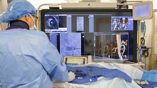
Our interventional radiologists are specialists in minimally invasive techniques. In addition to the training that all radiologists receive, these specialists have advanced fellowship training in interventional radiology, plus extensive real-world experience.
Our program is accredited by the International Accreditation Commission, which ensures high-quality care for our patients.
Our team of interventional radiologists and physician assistants coordinates patients’ complete care – from imaging evaluation to post-procedure follow-up – maintaining a high level of communication with each patient throughout the process.
In addition, we coordinate closely with experts from across the UT Southwestern community.
Our Noninvasive Vascular Imaging Services
The Comprehensive Noninvasive Vascular Imaging Laboratory uses state-of-the-art imaging technology to diagnose vascular diseases and assist our cardiologists, cardiothoracic surgeons, and vascular surgeons with diagnosing and planning surgical procedures.
These imaging studies provide important information required to plan procedures such as aortic aneurysm repair and transcatheter aortic valve repair, a new method of replacing the aortic valve without requiring open-heart surgery.
We use a variety of medical examinations to accurately diagnose patients’ vascular conditions. These examinations include:
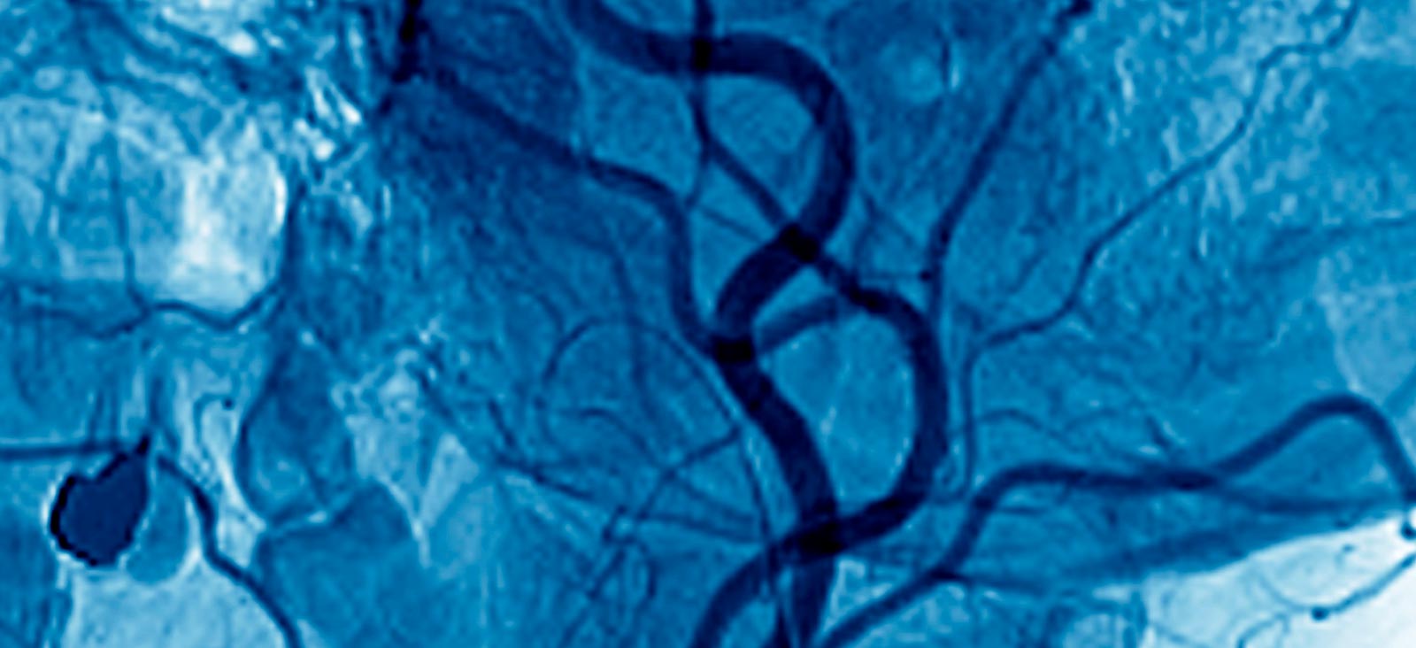
- Computed tomography angiography (CTA): CTA is a noninvasive exam that uses computed tomography (CT) technology to look at the blood vessels. It is used to evaluate abnormal narrowing or enlargement of blood vessels in the chest, abdomen, pelvis, and extremities. Our CTA techniques minimize radiation exposure while providing high-quality diagnostic images.
- Magnetic resonance angiography (MRA): MRA is a noninvasive exam that uses magnetic resonance (MR) technology – not ionizing radiation – to look at blood flow through the blood vessels. MR angiograms are interpreted by diagnostic radiologists who are fellowship trained in MR imaging.
- Vascular ultrasound: This noninvasive exam uses high-frequency sound waves to create images to detect the rate of blood flow through blood vessels. Ultrasound imaging can be used to detect blood clots and narrowed blood vessels throughout the body. It can be used to evaluate varicose veins and determine candidates for endovenous (vein) ablation.
Related Conditions and Treatments

Search for opportunities to participate in a clinical research study.
We’re one of the world’s top academic medical centers, with a unique legacy of innovation in patient care and scientific discovery.

MedBlog