Ocular Oncology

Diagnosis of Eye Tumors

Appointment New Patient Appointment or 214-645-2020

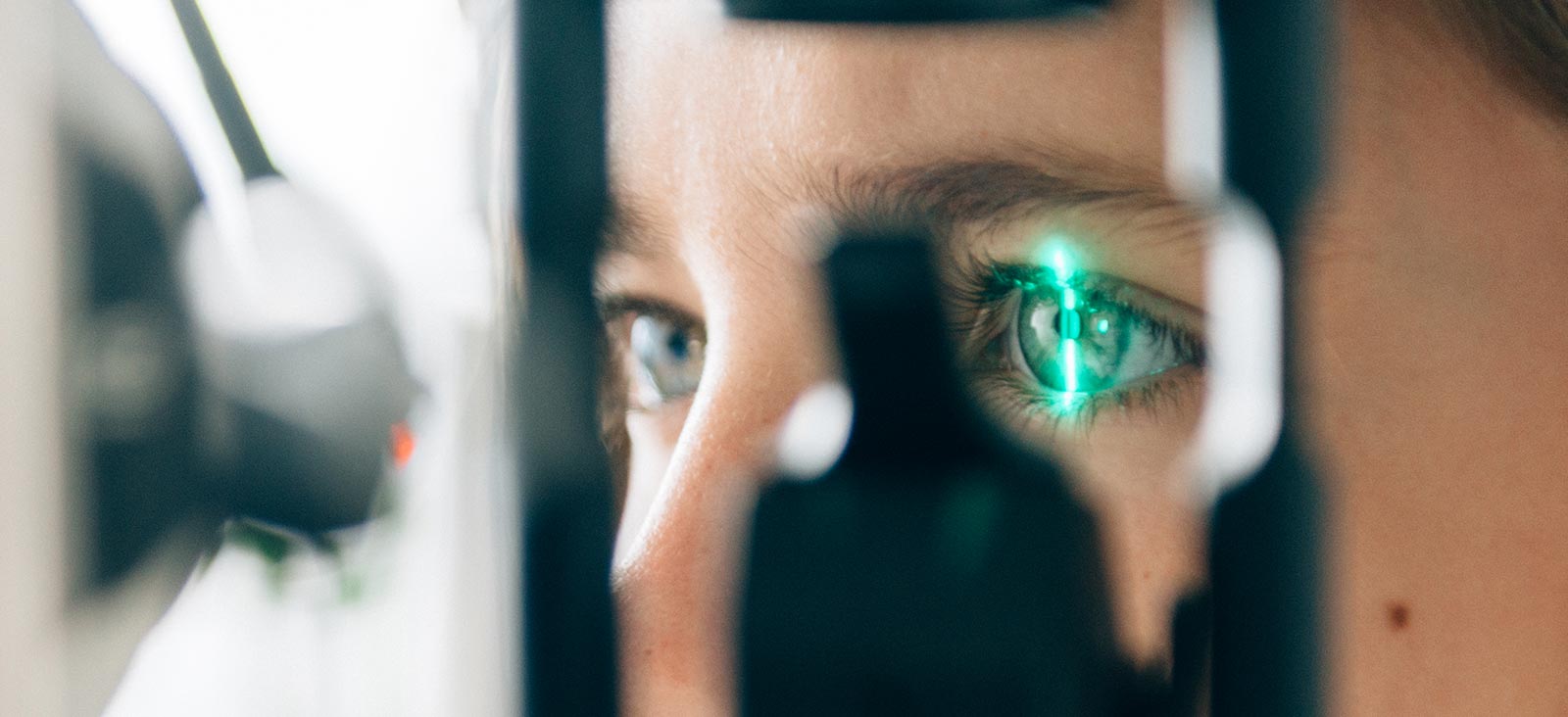
UT Southwestern’s ocular oncology team excels at diagnosing eye tumors and determining an individualized course of treatment for each patient. We may use ophthalmic imaging, which focuses on the eye, or systemic imaging, which involves other parts of the body.

Ophthalmic Imaging

To diagnose an eye tumor, numerous tests using advanced ophthalmic imaging equipment are often required.

These tests may include:

  • Fluorescein angiography: A procedure in which fluorescent dye is injected into a vein in the arm or hand to illuminate blood vessels in the retina. Photographs of the eye are taken using a special camera that can detect fluorescence. This test is especially helpful in evaluating abnormal blood vessels.
  • Indocyanine green angiography: Like fluorescein angiography, this test (often called ICG) uses a special dye to illuminate blood vessels and specifically evaluates deeper blood vessels located in the choroid, which is the vascular tissue layer behind the retina.
  • Optical coherence tomography (OCT): This test creates high-resolution scans of the retina or cornea using lightwaves. OCT is extremely useful, and it is quick and easy for the patient. OCT is one of the most frequently used tests in ophthalmology, both in general and in ocular oncology in particular.
  • Ophthalmic ultrasound: This test is performed by a highly skilled specialist and uses sound waves to produce images of the eye and surrounding structures. It’s extremely helpful in evaluating tumors, retinal detachments, hemorrhage, and other abnormalities. For this test, the eye is numbed with drops and a probe is gently placed on the eye.
  • Photography: The most common imaging test in ocular oncology is photography, which is used to document and measure eye tumors and to monitor them for change over time. A specially trained ophthalmic photographer uses different types of cameras to take images of various parts of the eye, including the lid, cornea, iris, retina, and optic nerve.

Systemic Imaging and Testing

Patients with eye tumors often require imaging of other parts of the body to evaluate for the presence of other tumors or abnormalities, and they may require genetic testing to evaluate for conditions that can affect other parts of the body.

  • Systemic imaging studies: Imaging studies such as computed tomography (CT), magnetic resonance imaging (MRI), and positron-emission tomography (PET) may be recommended in certain patients. While such tests are rarely required to diagnose the eye tumor, they are often helpful in determining the extent of involvement of other parts of the body.
  • Genetic testing: Some types of eye tumors can be associated with genetic mutations (damage to genes in the DNA) that can be detected from a source of normal tissue, such as blood, sputum, or a swab of the inside of the cheek (buccal swab). Such testing may be coordinated with a visit to a genetic counselor at UT Southwestern.