Full Spectrum of Diagnostic Services
Diagnostic X-ray, or radiography, is a special method for taking pictures of areas inside the body. A machine focuses a small amount of radiation on the area of the body to be examined. The X-rays pass through the body, creating an image on film or a computer display.
The equipment, staff, and steps involved are different for each type of diagnostic X-ray procedure. However, they are all invaluable tools in detecting abnormalities and making early diagnosis of disease or injury.
UT Southwestern offers experience and expertise in all types of diagnostic X-rays, including technologies and techniques that might not be available at other medical facilities.
Conditions We Diagnose With X-Ray
Various types of diagnostic X-ray procedures are ordered for different reasons. Common procedures include:
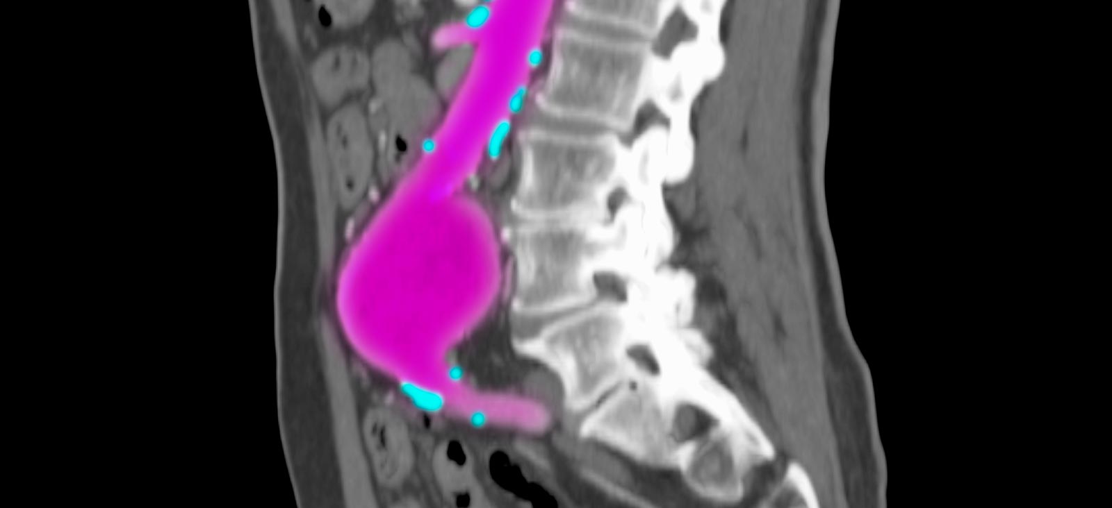
- Angiography: Uses an injection of contrast medium to image blood vessels in a specific part of the body. Angiograms show the function of blood vessels in the heart, lungs, kidneys, brain, arms, and legs.
- Arthrogram: Uses an injection of contrast medium into a joint. This procedure shows injury or disease in joints, arms, and legs.
- Upper GI (gastrointestinal) series: Uses a barium solution as a contrast medium and helps evaluate the function of the esophagus, stomach, and upper small intestine.
- Lower GI series: Uses a barium enema to evaluate the colon and rectum.
- Intravenous pyelogram (IVP): Uses a contrast medium injection to evaluate the kidneys, ureters, and bladder.
- Mammography: Uses a special X-ray machine to create images of breast tissue for detection of abnormalities.
Diagnostic X-Ray: What to Expect
Most routine X-rays do not require patients to prepare for the exam. However, special studies, such as contrast radiography or barium enemas, require patients to follow special instructions from the doctor.
Patients might be asked to make dietary changes leading up to the time of the exam. Patients might also be asked to leave jewelry at home, along with other metal objects that could interfere with the X-ray images. Patients might be asked to avoid using deodorants, body powders, or creams on the day of the appointment.
Patients will follow a different process depending on the diagnostic X-ray procedure. They might be asked to change into a gown or smock.
At the appointment, patients will meet X-ray professionals who are specially trained to help with the procedure:
- A radiologist is a doctor who specializes in imaging the human body.
- A radiologic technologist is trained to operate the equipment and obtain X-ray images.
- A radiologic nurse monitors vital signs, administers medication, and provides patient care during the procedure.
Once a patient has changed into the smock or gown, a technologist will escort him or her into the X-ray room to stand, sit, or lie on a table that is near an X-ray machine. An apron or shield might be placed over the patient’s body to protect sensitive organs during the exam.
The machine will take several X-rays, and the patient might be asked to adjust position during the test. It is important to remain still during each examination.
Patients might be asked to wait until the radiologist reviews images to be sure additional images aren’t needed. If the patient ingested a contrast medium or barium, it is important to drink plenty of liquid over the 24 to 48 hours following the scan to help pass the material.
The radiologist will review the images and send a report to the doctor, who will notify the patient of any findings. Patients can also request to receive images on CD.
Risks
While diagnostic X-ray procedures are generally safe and very effective, there is some exposure to radiation. However, the benefits of early detection and treatment far outweigh the risks.
Some people have an allergic reaction to contrast media, which is used in certain diagnostic X-rays. Symptoms of an allergic reaction include:
- Hives
- Itchiness
- Nausea
- Shortness of breath
- Weakness
Report these symptoms to the doctor, radiologist, or imaging technologist immediately. Tell the technologist if the patient has any known allergies to contrast media or iodine.
Please tell the doctor if the patient has had a reaction to contrast medium in the past or if the patient might be pregnant, because exposure to radiation can cause birth defects.
If patients have questions about a health condition that could affect the exam, please talk to a technologist or radiologist.


