In the quest for improved concussion care, a simple but advanced tool could give us hours of advance notice in diagnosing concussion and the complications that could arise in the days after the injury – all at the push of a button.
We already know that the NeurOptics NPiⓇ-200 Pupillometer device can give doctors an early warning about insufficient blood flow to a patient’s brain (cerebral ischemia), as well as brain swelling and other severe issues that can develop in the days after a hemorrhagic stroke.
Data from a UT Southwestern study of 56 patients showed that the pupils changed in seven of the 12 patients who developed cerebral ischemia after such a stroke. Pupil changes were detectable up to eight hours earlier than traditional methods of assessment; this early warning might grant us hours instead of minutes to intervene and reduce the risk of long-term brain deficits and cognitive loss.
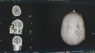
Today, UT Southwestern houses the world’s largest database for pupillometer research in acute brain injury. Our intensive care unit (ICU) uses the device on every patient after brain surgery and every patient with stroke, hemorrhage, or severe concussion/injury. Ongoing research will determine whether pupillometry might provide early warning for doctors to provide lifesaving interventions after concussions and other traumatic brain injuries.
How does pupillometry work?
Just as a speedometer measures changes in speed, a pupillometer measures changes in the pupil. The NeurOptics NPi-200 Pupillometer is a simple and user-friendly device. We hold the device in front of a patient’s eye and press a button to activate the measurements. A high-speed camera records how the pupil responds to light stimulus and computerized algorithms calculate the Neurological Pupil Index (NPi) in a matter of seconds.
The NPi is based on several measurements, including the pupil size and how fast the pupil responds to the light stimulus. Patients receive an NPi score on a scale of 0 to 5, with 3 to 5 considered a normal reading. Research suggests that an NPi below 3 could be a red flag that a patient might be experiencing a neurological complication.
What is the future of pupillometry?
Pupillometry research has vast potential to impact patient care. While currently only used in hospitals by doctors and nurses, we envision that the device could someday be used by coaches and trainers on the field of play.
For example, a father might notice that his daughter “isn’t acting right” after a hard body or head blow during a soccer match – parents know their children better than coaches do. Pupillometry could become part of the sideline concussion exam and offer measurable data to make safer, healthier decisions about return to play that would reduce long-term concussion symptoms after sports injuries.
Related reading: When should a student-athlete see a doctor for a concussion?
Particularly with neurological conditions such as concussion and aneurysm, early diagnosis and intervention is critical in reducing long-term damage. Pupillometry, while simple in theory, can potentially help reduce the risk of severe brain injury and even death.
Data from our pupillometry research reinforces the fact that doctors should consider the whole patient and not just the affected area of the body. A patient’s personal characteristics and seemingly unrelated physical reactions should be considered in neurological examinations, particularly in the days and weeks after an injury.
If you think that you or a loved one is experiencing concussion symptoms and need to see a doctor, call 214-645-8500 or request an appointment online.