
Search for opportunities to participate in a clinical research study.
Nuclear Medicine
New Patient Appointment or 214-645-8300
UT Southwestern Medical Center’s nuclear medicine services allow physicians and specialists to make accurate diagnoses and prescribe targeted treatments and therapies for a wide range of medical conditions, from the simple to the complex.
Our radiology team performs more than 800,000 inpatient and outpatient exams every year. We specialize in advanced technologies and the latest clinical innovations in today’s changing field of medical imaging.
Providing Valuable Insights into Structure and Function
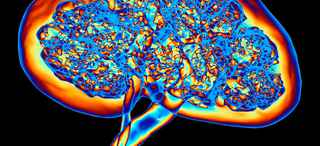
Nuclear medicine is a safe and effective method of studying how organs and other body parts function. A trace amount of radioactive material, called a radiopharmaceutical, is delivered to an area of the body and emits a form of radiation called gamma rays. These emissions are detected by a machine called a gamma camera, which converts them into images.
Nuclear medicine imaging is unique because it provides doctors with information about both the structure and the function of an organ, such as blood flow, filtration, and metabolism. It’s a way to gather medical information that would otherwise be unavailable, require surgery, or necessitate more expensive diagnostic tests.
A nuclear medicine exam is like an inside-out X-ray, in that the medicine records the radiation emitting from the body rather than radiation that is directed through the body.
The exam allows doctors to evaluate organs, and areas within organs, that aren’t evident on conventional X-rays. For example, instead of viewing just a detailed image of the kidney, a doctor can use nuclear medicine to see how the kidney is functioning.
UT Southwestern nuclear medicine specialists are highly trained and experienced in nuclear medicine tests and treatments. Our services include advanced imaging tools and techniques, many of which are not available at other medical facilities.
Conditions
More than 100 different nuclear medicine examination techniques allow doctors to evaluate organ function, making it an invaluable tool in early detection and diagnosis of many diseases.
Various types of nuclear medicine exams are given for different reasons:
- Bone scans can detect bone tumors, breaks, infections, and arthritis.
- Brain scans are done for people who have had a stroke or have seizures or related conditions.
- Cardiac scans can detect heart attacks and evaluate damage, measure the strength and efficiency of blood flow, and aid in stress tests.
- Gallbladder scans help detect abnormalities of biliary function.
- Lung scans can help detect problems such as blood clots, providing information about oxygen and blood supply.
- Renal scans can detect kidney tumors, cysts, obstructions, and other problems.
- Thyroid uptake scans give important information about the thyroid gland in the neck.
Types of Nuclear Medicine
Common procedures include:
- Stationary gamma cameras are positioned as close to the body as possible. Patients will stand, sit, or lie down, depending on the area to be scanned.
- SPECT, or single photon emission computed tomography, uses a rotating gamma camera that can move during the scan to provide a more complete picture of a particular body part.
- PET, or positron emission tomography, requires patients to lie on a table during the scan. The table moves through a tube, similar to the process of an MRI.
Nuclear Medicine: What to Expect
Preparation for the exam will vary according to what area is being tested. Our doctor will give patients special instructions prior to the appointment. Please follow them closely.
Patients might be asked to fast for several hours before the scan. For some scans, the radiopharmaceutical is administered hours or even days prior to the imaging portion of the scan.
We ask that patients wear comfortable, loose-fitting clothing, such as a sweatshirt without zippers or snaps. Patients might be asked to change into a gown, depending on the area of the body to be scanned.
When the patient arrives on the day of the appointment, he or she will meet a technologist who will conduct the nuclear medicine exam. The technologist will ask the patient to lie down or sit on a scanning table.
Our nuclear medicine technologist will administer the radiopharmaceutical in the form of an injection or by mouth in a liquid or capsule. Most scans require several different positions to capture several different views of the body. It is important that patients remain as motionless as possible during each scan.
Most of the low-level radiopharmaceutical will be excreted by the body naturally over the next 24 to 48 hours. Patients should drink plenty of water to help clear the material more quickly. Patients do not need to avoid contact with others during this time, but the doctor might suggest some precautions, such as flushing the toilet twice after use to reduce radiation exposure in the household.
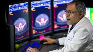
The nuclear medicine physician will review the images and send a report to the doctor, who will notify the patient of any findings. The patient can also request to receive images on CD.
Risks of Nuclear Medicine
It is important to note that while the nuclear medicine scan itself is a painless exam, it does involve exposure to radiation. However, the benefits of an accurate and early diagnosis far outweigh the risk.
The radioactive materials used in the exam are given in tiny amounts with exposure similar to what a patient would receive with a standard X-ray. They also lose their radioactivity very quickly and pass through the body usually within 24 to 48 hours.
If a patient is pregnant or breastfeeding, please alert the technologist. Exams using injected radioactive material are usually not recommended for women who are pregnant or breastfeeding.
A nuclear medicine physician or technologist can answer any questions a patient might have about a health condition that could affect the exam.
Related Conditions and Treatments


We’re one of the world’s top academic medical centers, with a unique legacy of innovation in patient care and scientific discovery.

MedBlog
Results: 2 Locations
Positron Emission Tomography (PET) Imaging Facility
at Bill and Rita Clements Advanced Medical Imaging Center 2201 Inwood Road, 2nd FloorDallas, Texas 75390 214-645-9729 Directions to Positron Emission Tomography (PET) Imaging Facility at Bill and Rita Clements Advanced Medical Imaging Center, Dallas Parking Info for Positron Emission Tomography (PET) Imaging Facility