Fetal echocardiogram: What to expect
August 7, 2025


Learning that your baby may be born with a heart condition is an emotional experience. And it’s one I’ve experienced as a pediatric cardiologist and as a mother.
When I was pregnant with my son, my doctor suspected he might have a heart abnormality. I had a fetal echocardiogram, which is a sophisticated ultrasound exam that helps determine whether a baby might be born with a congenital heart disease. The fetal echo gave us information to prepare for the surgery he’d need after delivery, and thankfully, he is happy and healthy today.
Heart problems are the most common type of birth condition, with about 1 in 100 babies born with a congenital heart disease such as a hole in the heart, a problem with the valves that control blood flow, or a heart rhythm condition. About 25% of babies born with a critical congenital heart disease will need surgery or another intervention by their first birthday.
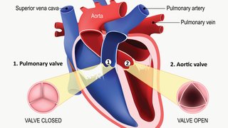
Compared with traditional ultrasound, a fetal echocardiogram gives us a clearer view of the fetal heart structure, heart rhythm, and blood flow. The exam is safe and noninvasive for you and your baby – it is performed with a wand on the belly like a regular ultrasound. The images and video from the exam will help guide your baby’s future care.
Let’s discuss why you might need a fetal echo, what to expect during the visit, and how we support you during and after the exam.
Why might I need a fetal echocardiogram?
Your pregnancy care provider may recommend this ultrasound based on fetal, maternal, or paternal criteria such as:
- Fetal anomalies: Your Ob/Gyn may see a possible heart problem during a routine pregnancy ultrasound.
- Family medical history: You or your partner might have family members with a congenital heart defect. Or prenatal carrier screening (genetic testing) showed a potential risk for a hereditary heart condition.
- Maternal health history: Some chronic health conditions in women such as diabetes, exposure to some medications, some types of thyroid disease, or autoimmune disorders such as lupus or Sjogren’s syndrome, can increase the risk of fetal heart problems. Conception by IVF (in vitro fertilization) also carries increased risk for congenital heart defects.
Most fetal echocardiograms are done between 18-24 weeks of pregnancy when we can get a clear picture of the structure and function of the baby’s heart. Often, fetal heart conditions happen sporadically, which means there is nothing you or your partner could have done differently to prevent it. Getting a fetal echo is the first step in understanding whether a congenital heart condition exists and giving you details to make informed decisions about the baby’s care.
Related reading: 4 things to consider before inviting guests to a baby’s ultrasound
What should I expect at my fetal echo appointment?
How to prepare
These tips can help you feel more prepared for your fetal echo:
- Write down your questions and concerns: That way, your care team can address what is important to you if the emotions become overwhelming.
- Bring a support person: Your partner or a close friend or family member can take notes and help you sort through the information.
- If you can, avoid bringing other children: You might get upsetting news during this visit, and you likely will want the space to express your emotions.
- Pack a drink and light snacks: Your fetal echo will take about an hour, and you may have downtime prior to being called to your exam room.
During the exam

You will first meet with a fetal nurse coordinator who will talk with you about your pregnancy, personal and family health history, and medications. In our particular unit, since the health and well-being of the mother is so critical to the well-being of the family, we also talk to each patient and ask questions to help determine whether you might have symptoms of anxiety or depression, which are common among patients who are pregnant. This allows us to provide helpful resources if concerns are noted.
Then the nurse will take you to the scanning room, which will be darkened to get the best images. Each scanning room has a large screen, so you and your support person can see the imaging in real time.
We’ll ask you to recline on the exam bed. A sonographer will apply a lubricant to your abdomen and use an external wand to conduct the ultrasound, focusing on the tiny structures that form your baby’s heart. A cardiologist may join you in the room to gather additional images. This ensures that your care team has a full picture of your baby’s heart before discussing the results.
You do not have to lie perfectly still for the hour-long exam. We may ask you to shift positions, and you can feel free to ask for a break. You will have access to medical interpreter services, if needed.
Getting the results
Typically, we discuss the findings at the end of your visit. In some cases, we need more information, and we may recommend another fetal echo later in your pregnancy.
You and your support person will meet with your care team to review the results and discuss what the findings might mean for your baby’s future health. Sometimes this conversation includes difficult news. Your care team will be there to support you through one-on-one counseling from a congenital heart disease expert and other specialists such as a maternal-fetal medicine doctor, psychologist, or social worker. We will provide a safe environment to process your feelings and receive honest, compassionate answers to all your questions.
During your pregnancy and after giving birth if there is a particularly severe cardiac abnormality or a cardiac abnormality in addition to other findings that is not amenable to medical or surgically curative interventions, we also provide access to palliative care services that help you to prepare for your baby’s future care, which may include discussion of which procedures are desired and not desired as well as resources to support your family in managing anxiety or grief if needed.
“Congenital heart defects can happen sporadically, with a majority of cases having no clear genetic or environmental cause. I always encourage parents to release any feeling of guilt as best as they can.”
Maria Ossa Galvis, M.D.Assistant Professor of Pediatrics, UT Southwestern
Making a plan for your baby’s heart care
Your care team will recommend a plan based on the fetal echo findings and what matters most to you and your family. For most women, pregnancy care doesn’t change except for possibly more ultrasounds and clinic visits.
Some congenital heart conditions resolve on their own without intervention. One example of this is a small hole in the heart; it sounds serious, but it is common and often closes later in the pregnancy or even after birth. In these cases, we’ll recommend observation and possibly a follow-up echocardiogram. If your baby has a less severe heart condition, the newborn can stay with you in the birth suite after delivery.
Babies with severe congenital heart conditions may require medication as well as one or more surgeries in their first year of life. In some of these cases, delivery hospital location plans may need to change to provide the best care for your baby. Typically, mode of delivery decisions are not impacted by fetal heart anomalies, but talk with your own doctor about whether C-section or vaginal delivery would be safest for you and your baby.
You will want to choose a delivery hospital with a neonatal intensive care unit (NICU) and a neonatal cardiologist on staff. Both William P. Clements Jr. University Hospital and Parkland Health are Level III NICUs staffed by UTSW neonatologists and cardiologists. At Clements, all families get a private room that includes a comfortable chair, couch, sink, and TV so you can feel a little more at home while we care for your baby. Extended family members can virtually visit the baby through our secure NICU camera system. Parkland Health’s high-volume NICU cares for more than 1,300 newborns a year. On average, the team takes care of about 100 newborns each month.
Some babies need to be transferred to Children’s Health, which has both a Level IV NICU and a dedicated cardiac intensive care unit. Experts are available 24/7 to care for babies with even the most complex heart conditions.
As a parent who has been through this experience, I understand how challenging it can be to wait for and process the results of a fetal echo. Detecting heart problems before a baby is born provides an opportunity to be prepared for delivery and what comes after the baby is born. You won’t go through it alone – your care team will be at your side to help you prepare the best you can. Together, we’ll make a plan based on what is most important for you and your family.
Catherine Ikemba, M.D., a fetal cardiologist and Professor of Pediatrics at UT Southwestern, contributed to this report.
To talk with an expert about your pregnancy, make an appointment by calling 214-645-8300 or request an appointment online.