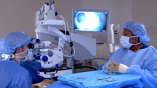
At UT Southwestern Medical Center, our skilled ophthalmologists have experience in treating all kinds of eye diseases and conditions, including all types of glaucoma. Our ophthalmic specialists offers patients the latest diagnostic techniques and treatment options to slow the progression of the condition and preserve vision.
What Is Glaucoma?
Glaucoma is a group of diseases that affect the optic nerve, which transmits images from the eye to the brain. Glaucoma often results from high pressure inside the eye (intraocular pressure). The condition causes vision loss and can lead to blindness.
Expert Care for All Types of Glaucoma
UT Southwestern offers patients individualized, leading-edge treatment for glaucoma to slow its progression and preserve vision. Our glaucoma specialists have access to the latest diagnostic technologies, such as Humphrey visual field analyzers and optical coherence tomography (OCT). We offer specialized treatment such as traditional glaucoma surgery (trabeculectomy and drainage implant surgery), combined small-incision cataract/glaucoma surgery, and minimally invasive glaucoma surgery, or MIGS (including iStent®, Cypass®, and the Kahook Dual Blade®).
Types of Glaucoma
In a healthy eye, a clear fluid (aqueous humor) fills the front of the eye and normally flows in and out through channels where the iris and cornea meet. Because glaucoma often results from high intraocular pressure, anything that blocks or slows the flow of this fluid increases pressure.
The major types of glaucoma are:
- Open-angle glaucoma: More than 90 percent of adults with glaucoma have this type. In open-angle glaucoma, fluid inside the eye pressure increases slowly over a long time, and patients often do not notice a change. As pressure on the optic nerve increases, it can damage the nerve and cause vision loss such as blind spots.
- Angle-closure glaucoma: Also known as closed-angle glaucoma, this type occurs when the fluid suddenly becomes blocked, leading to a quick, dramatic pressure increase inside the eye. Angle-closure glaucoma usually causes immediate, noticeable symptoms such as severe eye pain and blurry vision. This type of glaucoma is a medical emergency and requires immediate treatment to prevent blindness.
- Low- or normal-tension glaucoma: Some people with normal eye pressure can develop this type of glaucoma, a form of open-angle glaucoma.
- Congenital glaucoma: Some babies are born with a defect in the eye that slows the normal fluid drainage. Congenital glaucoma usually causes noticeable symptoms and signs very early, and prompt surgery provides children with an excellent chance for good vision.
- Secondary glaucoma: Either open- or closed-angle glaucoma can develop as a result of a known cause, such as another eye disease, a medical condition, an injury, or certain medications. Some types of secondary glaucoma include:
- Pigmentary glaucoma: In younger, nearsighted patients, pigment from the iris can dislodge and clog the eye’s internal drainage channels, leading to build up of pressure inside the eye and a secondary glaucoma.
- Traumatic glaucoma: Eye injuries can damage and block the fluid drainage system in the eye, which increases eye pressure and can lead to glaucoma. Traumatic glaucoma can develop immediately after an injury or even years later, so ongoing monitoring is important.
Causes and Risk Factors for Glaucoma
The causes of open-angle glaucoma are unknown. Causes for other types of glaucoma include:
- Dilating eye drops
- Certain medications such as corticosteroids
- Eye diseases
- Other conditions such as diabetes
- Previous eye injury or eye surgery
- Congenital (present since birth) defect
Several factors can increase a person’s risk of developing glaucoma, such as:
- Advanced age
- Extreme nearsightedness
- African or Hispanic ancestry
- Extremely low blood pressure
- Diabetes (in some cases)
- Family history of glaucoma
- Thin cornea (clear layer covering the front of the eye)
Preventing Vision Loss
Routine dilated eye examinations with an eye specialist are the best way to detect glaucoma and can help prevent vision loss. The American Academy of Ophthalmology recommends having an eye examination:
- Every 5 to 10 years for people under age 40
- Every 2 to 4 years for people ages 40 to 54
- Every 1 to 3 years for people ages 55 to 64
- Every 1 to 2 years for people age 65 and older or who have glaucoma risk factors
Glaucoma Symptoms
Early detection and treatment by an ophthalmologist can prevent vision loss. The most common type of glaucoma, open-angle, often shows no symptoms at first, which is why it’s important for patients to have regular eye exams with a specialist.
Symptoms of glaucoma can include:
- Slow loss of peripheral (side) vision
- Sudden, severe pain in one eye
- Reduced or cloudy vision
- Nausea or vomiting
- Halos around lights
- Redness or swelling of the eye
- Sensitivity to light
- Cloudy appearance in the front of the eye
Diagnosing Glaucoma
UT Southwestern eye specialists can diagnose glaucoma through a comprehensive eye exam. The exam might include several of the following tests:
- Vision tests, including automated visual field tests to check peripheral (side) vision
- Dilated eye test to examine inside the eye
- Tonometry, a painless test that measures pressure inside the eye
- Slit-lamp exam to look at structures inside the eye, such as the retina, lens, cornea, iris, and anterior chamber
- Gonioscopy using a special lens to check the drainage angle for abnormalities
- Imaging, such as fundus photography, of the inside of the eye to examine the optic nerve and measure any changes over time
- Indirect ophthalmoscopy to evaluate the peripheral retina
Eye pressure is measured in millimeters of mercury (mm Hg). Normal eye pressure typically ranges from 10 to 22 mmHg. Pressure higher than 22 mmHg can indicate an increased risk for developing glaucoma. However, glaucoma can develop even when eye pressure measures in the normal range.
Treatment for Glaucoma
The treatment goal is to reduce eye pressure to avoid damage to the optic nerve. Treatment options vary by the type of glaucoma and include:
- Application of daily medicated eye drops to reduce pressure by helping fluid drain from the eye or reducing the amount of fluid the eye produces
- Pills to reduce eye pressure (less common)
- Laser surgery to open the channels for the fluid inside the eye to drain more easily
- Minimally invasive glaucoma surgery (MIGS), which offers less invasive surgical options to lower pressure inside the eyes; it’s typically done in conjunction with cataract surgery
- Traditional surgery, such as trabeculectomy, to create an opening in the eye for fluid to drain more easily to an external bleb or reservoir