
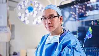
Combining attentive, compassionate care with our extensive clinical and research resources, UT Southwestern's cardiology experts and vascular specialists deliver individualized care within pre-eminent healthcare facilities.
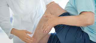
Varicose Veins
New Patient Appointment or 214-645-8300
Vascular experts and interventional radiologists at UT Southwestern Medical Center provide safe and effective treatments for varicose veins and spider veins.
Specializing in minimally invasive, targeted treatments, our team treats the source of the disease rather than just the symptoms. We offer all available varicose vein treatments under one roof, including the latest, most advanced ablation equipment.

Eliminating Discomfort and Pain, Improving Appearance
Varicose veins are enlarged veins that are visible through the skin and can appear as blue or purple, twisted, knot-like cords. Varicose veins can occur anywhere in the body but are more commonly found on the legs. This condition is caused by increased blood pressure inside the superficial leg veins.
When veins are healthy and working properly, they return blood to the heart for re-oxygenation, but when valves in the veins fail – or become damaged or leak – some of the blood flows backward rather than toward the heart, accumulating in the veins and causing them to thicken, elongate, and sometimes bulge.
Left untreated, varicose veins can cause serious health issues such as venous leg ulcers, skin discolorations, or blood clots. They affect about 20% of the U.S. adult population and half of those age 50 and older.
For many patients, procedures to correct varicose veins are necessary to decrease or eliminate discomfort, aches, and pains. For others, treating varicose and spider veins is important for cosmetic reasons.
Symptoms of Varicose Veins
Varicose veins are common in patients with a family history of the condition and in women during or immediately following pregnancy. Symptoms include:
- Discomfort
- Achiness, heaviness, and fatigue in the affected area
- Itchiness or numbness
- Swelling
Progression of varicose veins can lead to:
- Skin discoloration
- Leg ulcers
- Blood clots
Treating varicose veins early – with minimally invasive, nonsurgical procedures – can prevent these progressive symptoms.
Diagnosis
Physicians might request a combination of diagnostic procedures to confirm varicose veins. Some of these procedures include:
- Duplex ultrasound: A type of vascular ultrasound procedure used to assess blood flow and the structure of the leg veins. Duplex means that two modes of ultrasound are used – Doppler and B-mode. The B-mode transducer (like a microphone) obtains an image of the vessel being studied. The Doppler probe within the transducer evaluates the velocity and direction of blood flow in the vessel.
- Color-flow imaging: Also called triplex ultrasound, this procedure is similar to duplex ultrasound but uses color to highlight the direction of blood flow. Vessels in which blood is flowing are colored red for flow in one direction and blue for flow in the other direction, with a color scale that reflects the speed of the flow.
- Magnetic resonance venography (MRV): A diagnostic procedure that uses a combination of a large magnet, radiofrequencies, and a computer to produce detailed images of organs and structures within the body. An MRV uses magnetic resonance technology and intravenous (IV) contrast dye to visualize the veins. Contrast dye causes the blood vessels to appear opaque on the X-ray image, allowing the physician to visualize the blood vessels being evaluated. MRV is useful in some cases because it can help detect causes of leg pain other than vein problems.
Treatments for Varicose Veins
At UT Southwestern, our vascular specialists and interventional radiologists offer surgical and nonsurgical treatment for varicose veins.
Nonsurgical Methods
- Ablation: Involves the insertion of a thin, flexible tube called a catheter into a varicose vein. The tip of the catheter heats the walls of the varicose vein and destroys the vein tissue. Once destroyed, the vein is no longer able to carry blood and is absorbed by the body.
- Compression stockings: These elastic stockings squeeze or compress the veins and prevent blood from flowing backward. Compression stockings can help with healing of skin sores and prevention of additional sores. They are effective in treating varicose veins if worn daily and can prevent the need for more invasive treatment.
- Elevation of the legs: Elevating the feet above the heart three or four times a day for about 15 minutes at a time can help. Flexing (bending) the legs occasionally can also help keep blood circulating.
- Laser treatment: Until recently, laser treatment was mainly used to treat spider veins on the face. However, newer laser technology can now effectively treat varicose veins as well. The physician inserts a tiny fiber into a varicose vein through a catheter. The fiber sends out laser energy that destroys the diseased portion of the varicose vein. The vein closes and the body eventually absorbs it.
- Microphlebectomy: This minimally invasive procedure removes the varicose vein via a tiny nick made with a special set of tools.
Surgical Methods
- Small incision avulsion: This procedure involves passing hooks through small incisions and can be done alone or together with vein stripping.
- Transilluminated-powered phlebectomy: This vein-removal procedure uses a bright light to illuminate the vein. A device is passed through a tiny incision and removes the vein with suction.
- Vein stripping: This procedure involves tying off all varicose veins associated with the leg's main superficial vein and removing it from the leg. Removing veins from the leg will not affect blood circulation in the leg because deeper veins take care of the increased blood circulation.
Related Conditions and Treatments

Search for opportunities to participate in a heart or vascular research study.
We’re one of the world’s top academic medical centers, with a unique legacy of innovation in patient care and scientific discovery.

Results: 4 Locations
West Campus Building 3
2001 Inwood RoadDallas, Texas 75390 214-645-8300 Directions to West Campus Building 3, Dallas Parking Info for West Campus Building 3
Surgical Specialties
at UT Southwestern Frisco 12500 Dallas Parkway, 2nd FloorFrisco, Texas 75033 469-604-9150 Directions to Surgical Specialties at UT Southwestern Frisco, Frisco Parking Info for Surgical Specialties
Vascular Medicine
at UT Southwestern Medical Center at RedBird 3450 W. Camp Wisdom RoadDallas, Texas 75237 214-214-5810 Directions to Vascular Medicine at UT Southwestern Medical Center at RedBird, Dallas Parking Info for Vascular Medicine
Vascular Medicine
at UT Southwestern Medical Center at Coppell 2999 Olympus Blvd., 3rd FloorCoppell, Texas 75019 469-647-4446 Directions to Vascular Medicine at UT Southwestern Medical Center at Coppell, Coppell Parking Info for Vascular Medicine