What is a brain AVM? One patient’s journey through diagnosis and treatment
August 19, 2026


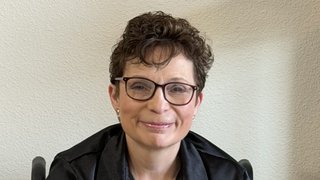
At age 49, Sharron Fowler felt as if she was in the prime of her life. A nurse by trade, she devoted herself to her work each week and looked forward to one daughter’s upcoming wedding and another’s soon‑to‑arrive granddaughter. She didn’t have time to be sick.
But in 2012, when she noticed her balance and cognitive function was off, she began to worry it was more than a pulled muscle or simple brain fog. She was admitted to a hospital for observation and testing, and discharged a few days later with no clear diagnosis.
As she continued to struggle with her balance, she spent eight more days in a hospital and faced a range of frightening possibilities – from altered mental status to multiple sclerosis to cancer. Sharron insisted on being transferred more than 100 miles away to UT Southwestern, following a friend’s recommendation.
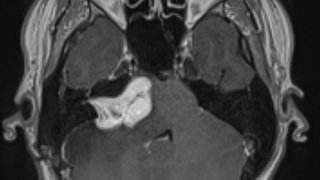
Upon admission, Babu G. Welch, M.D., Medical Director of the Neurosurgery Ambulatory Clinic and Director of the Microvascular Laboratory at UT Southwestern’s Peter O'Donnell Jr. Brain Institute, reviewed her record. After more testing, they finally had an answer – a confirmed arteriovenous malformations (AVM) of the occipital portion of Sharron’s brain.
What is an arteriovenous malformation?
AVMs form when arteries and veins develop in an abnormal way, creating a tangle of blood vessels around critical parts of the body, such as the brain or heart. When the blood vessels tangle this way, blood can flow faster than normal, bypassing the regular capillaries and affecting how oxygen is delivered to surrounding tissue.
In some cases, AVMs may not cause symptoms for years, while in others, they can lead to serious complications.
Around the brain, AVMs can lead to:
- Chronic headaches
- Seizures
- Stroke
- Bleeding or hemorrhage
- Vision changes or other neurological symptoms
AVMs can be difficult to diagnose

When Sharron was first diagnosed, she faced the same confusion that often surrounds AVMs. Affecting less than 10 per 100,000 people, they are rare, symptoms are unpredictable, and without imaging they are frequently overlooked. They have a slight predominance in women and typically surface between ages 30 and 50 — though they can also be present in children (UT Southwestern now has dedicated pediatric expertise for AVMs).
Compounding the challenge, many patients live for years without symptoms, unaware a potentially dangerous lesion is there at all. While many patients with AVMs experience symptoms such as headaches, seizures, or balance problems, some are discovered incidentally — for example, in patients with longstanding migraines who get imaging for the first time. Patients with clinical syndromes like hereditary hemorrhagic telangiectasia (HHT) may also have AVMs.
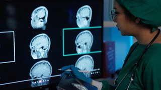
Through imaging and testing, specialists determine the AVM’s type, size, and location to guide the most appropriate treatment approach. While an MRI can identify AVMs, managing them after a diagnosis requires a coordinated plan between medical and surgical specialists. Because of this complexity, patients often seek care at referral centers such as UT Southwestern.
Individualized treatment for each patient
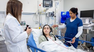
Once an AVM is detected, the cerebrovascular team at UT Southwestern works to determine the best path forward. This begins in a conference that occurs every week where all specialties discuss, and sometimes debate, the best plan for each patient. Every step is coordinated, and shared expertise arrives at an individualized plan rather than a one-size-fits-all approach.
Depending on each patient’s conditions, the plan might involve one or more of these treatments:
- Surgery: Having neurosurgeons directly access the AVM to remove it.
- Embolization: Performing a minimally invasive procedure where a catheter delivers medicine or synthetic substances to block blood flow to an AVM to shrink or close it off.
- Radiation: Using targeted radiation over time to close off the abnormal blood vessels without invasive surgery.
- Simple observation: Monitoring the AVM with regular imaging and follow-up visits when the risk of treatment may outweigh the benefits.

Sharron describes the relief and the reassurance she felt from the multidisciplinary team overseeing her case. “Dr. Welch introduced the team, and my room was filled with white coats around my bed,” she says. “I remember saying, ‘There’s no way you’ll all be back tomorrow — it’s New Year’s Day.’ But he smiled and said, ‘We’ll be here, won’t we, team?’ And in unison, they answered, ‘Yes, we’ll be here.’”
Throughout her time at UT Southwestern, Sharron was treated by a coordinated team of specialists.
“In this case, a neurosurgeon removed the AVM, and an interventional radiologist blocked off the abnormal vessels to the AVM (embolization) beforehand to make surgery safer,” Dr. Welch says. “A neurocritical care specialist helped with postoperative care, and neurorehabilitation was critical to her recovery.”
More than a decade after her surgery, Sharron continues living a healthy life, working as a nurse, and sharing her experience with others facing an AVM diagnosis. Years after her treatment, she and Dr. Welch reconnected at a medical conference where she spoke about her journey. It was a full-circle moment that reflected just how far she had come since treatment.
Related: Read "Most brain aneurysms aren’t ‘time bombs’: The right treatment may be nothing at all"
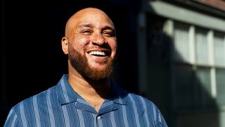
At UT Southwestern, we treat a large number of AVMs, and every case requires a true multidisciplinary approach. What really sets us apart is that we don’t begin treatment unless we have a clear, coordinated plan to see it all the way through.
Babu G. Welch, M.D.Medical Director of the Neurosurgery Ambulatory Clinic and Director of the Microvascular Laboratory at UT Southwestern
Living with AVMs
Being diagnosed with an AVM often goes beyond a single treatment. Blood pressure control, a healthy diet, and general wellness are recommended just like with anyone else. If seizures are present, standard seizure precautions apply.
“The greater challenge for many patients is the ‘what if.’ Living with an untreated AVM can create anxiety about bleeding risk, even when that risk is low,” Dr. Welch says. “Some patients are comfortable with careful monitoring. Others find the uncertainty overwhelming, and that psychological burden can factor into treatment decisions.”
The team at UT Southwestern believes that care must address both the biology of the lesion and the mindset of the person living with it.
For patients under observation, follow-up is individualized with imaging spaced out over years, especially if the AVM is stable. Patients are educated about symptoms that should prompt urgent evaluation, such as sudden severe headache or changes in seizure patterns. Over time, some patients become comfortable and simply check in periodically.
For those who undergo surgery like Sharron did, follow-up is more structured, and recovery can involve neurocritical care, rehabilitation, and, sometimes, neuropsychological support.
“There is no single pathway,” Dr Welch stresses. “Treatment decisions depend on the AVM’s size and location, its relationship to critical brain structures, how the patient presented, and how they are tolerating the diagnosis emotionally. A long-term partnership with an experienced team is essential.”
Related: Read "'I feel like I’ve been shot in the head’: Mother, daughter survive complex brain aneurysms"
Advanced care for arteriovenous malformations
Treatment of AVMs has continued to evolve since Sharron’s 2012 diagnosis:
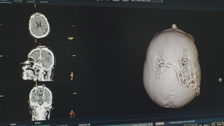
- Better imaging: Advances in imaging allow specialists to better understand what’s happening in the brain and how an AVM may affect movement, sensation, and vision. UT Southwestern is currently investigating the application of functional MRI to improve AVM outcomes, while researchers continue to explore new ways to use angiography to evaluate and treat AVMs.
- Minimally invasive techniques: Small catheters allow specialists to reach the arteries that feed the AVM with greater precision. Medications and treatment devices can be passed through these catheters to both improve understanding and treat the AVMs.
- Hybrid operating suites: Operating theaters called “hybrid suites” put everything the surgeon needs – surgical and radiology equipment – in one spot to decrease patient movement. UT Southwestern has utilized hybrid operating suites for surgical treatment of AVMs since 2007. Many major centers around the country such as Cleveland Clinic, University of Washington, and University of Pennsylvania have looked to this expertise to build their own facilities.
Life beyond an AVM diagnosis
Sharron’s experience highlights how AVM care extends beyond a single procedure and into long-term monitoring, recovery, and quality of life.

“We take pride in being a long-standing referral center for AVMs. There’s a deep culture here built around caring for patients with these complex lesions and helping them lead normal lives,” Dr. Welch says. “In every specialty involved, there are senior physicians who have been here for more than 20 years.”
Radiation oncology, neurosurgery, interventional radiology, neurology, neurocritical care, and neurorehabilitation — each area has experienced anchors who have contributed to the evolution of AVM care.
“Continuity matters,” Dr. Welch says. “It means we’re not just treating a moment in time — we’re following patients for years, sometimes decades, and learning every step along the way.”
For many patients, that long-term relationship involves follow-up care tailored to their individual needs, which may include:
- Regular imaging: MRI scans, angiograms, or other imaging studies may be used to monitor the AVM and track long-term outcomes.
- Rehabilitation services: Physical therapy, occupational therapy, speech therapy, and neurorehabilitation may help patients recover and regain function.
- Seizure management: Patients who experience seizures may require ongoing monitoring, medication management, and seizure precautions.
- Long-term specialist care: Follow-up visits allow patients and their care teams to monitor progress, review imaging results, and address concerns as they arise.
That long view strengthens care so that every case adds to what teams know, every follow‑up deepens understanding, and every recovery helps shape how the next patient is treated. The UT Southwestern team shares a commitment not only to guiding patients through treatment, but also to supporting their long‑term recovery and life well beyond their AVM.
To speak with an expert about AVM treatment options, make an appointment by calling 214-645-2300 or request an appointment online.