When I was in college, I fell in love with time-frequency analysis – studying data to pinpoint when and how often an event occurs. That’s right, I was one of the cool kids.
I was determined to become an electrical engineer but then, one fateful day in biology, my professor mentioned the brain emits electrical signals, too. And thus began my journey to becoming a MEG Scientist.
MEG technology (magnetoencephalography) is a revolutionary brain signal processing tool that allows doctors to retrieve an enormous volume of data from a patient’s brain with absolutely no pain, invasiveness, or emissions – it is safer than holding a cell phone against your head.
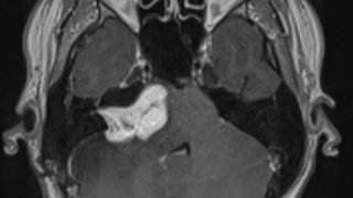
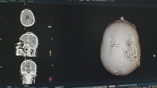
In just one second of testing, we can gather up to half a million data points. Each data point can be synced to a 3D model of a patient’s brain. The result? A map that gives us, within a millimeter’s precision, the origin of disruptive brain events such as epileptic seizures.
At UT Southwestern, senior post-doctoral research associate Amy Proskovec, Ph.D., and I harness the power of MEG technology to help improve epilepsy surgery outcomes, zeroing in on where in the brain a patient’s seizures originate. UT Southwestern offers the most advanced MEG technology currently available anywhere, and the only MEG in Dallas.
MEG technology guides the most precise epilepsy surgery – and it all begins with a color-changing ceiling, a Darth Vader helmet, and ’80s-style glasses.

Enter the MEG suite. Choose your color
Epilepsy can rob a patient of personal control in so many ways. Our goal is to help patients regain their lost autonomy, starting in the MEG suite.
On the ceiling in the prep area, we’ve installed a wavy lighting treatment. It’s meant for enjoyment only – there’s no medical reason behind it. But patients seem to enjoy the chance to set the color mood while we help them don their head attire and prepare for the exam.
Related reading: Who should consider epilepsy surgery, and when?

Dressing for the event, I mean, exam
First, we ask patients to put on a special cap – OK, it looks like a shower cap with 64 electrodes. Each of these electrodes measures electrical signals in the brain directly from the scalp. Sweet, sweet data gathering.
Next, it’s retro glasses time. Patients sit in a comfortable chair and put on bulky looking but lightweight glasses that help hold a receiver in place as we digitally record the location of each sensor in relation to their head and brain. We pass a digital wand that looks like an enlarged iPad stylus over each electrode on the cap, logging what we call the patient’s “head shape,” consisting of several 3D renderings of your head.
We’ll compare the 3D images to your MEG data readout to your MRI, creating what is essentially a Google Map of your brain signal activity. Neat, right? Now it’s time for the exam itself. On to the MEG room!
The sound of silence: Inside the MEG room
The MEG room has thick walls made of three layers of copper, aluminum, and a nickel-iron alloy called mu-metal. The world outside this room is full of electromagnetic “noise” from devices such as computers, and basically anything made of metal or moving parts. Humans can’t hear this noise, but the MEG can.
We use the thick doors to block out every other signal so the MEG can detect the brain’s magnetic field energy, which is measured on the Femto-Tesla scale – about 1 quadrillion times smaller than the signals emitted by the MRI machines in the next suite over and 10 billion times smaller than the Earth’s natural magnetic field. This is the noise we want to measure, and nothing else.
Inside the MEG room is a comfy chair that looks like a recliner. We’ll ask you to sit or lie down for an hour – you can take a nap if you want – while the MEG machine reads the noise signals from your brain.
To get the proper readout, the next step is to put on the MEG helmet, also known as Vader Time.

‘I am your doctor’
We call the MEG helmet a Darth Vader helmet because of its shape. It doesn’t cover the face, but it looks like something from the movie “Star Wars.”
The helmet is like a wearable brain scanner with 306 sensors. We have a mock helmet we let pediatric patients wear around the suite during prep so they can get used to it.
Once we connect the cap and helmet, the patient settles into the recliner and we explain how the exam works. Basically, the MEG helmet sensors, as well as the electrode cap, deliver precise brain signal readouts to the MEG computer.
Then, we do the only remotely startling step in the exam. We suction close the door. It’s not super loud – it’s quieter than slamming a door when you’re mad – but we warn patients that it might startle them because the room is so quiet. The door is shut to keep out those pesky magnetic noises from the nearby MRIs, computers, cars, etc. We can still see and hear you over the camera system. And, of course, there is a big red button that opens the door in an emergency if you get nervous.
The MEG is completely passive. It emits no radiation and is FDA-approved for use in newborn infants and pregnant women, so there's nothing to worry about in that regard.
That’s all there is to it, for the patient, anyway. Next up, it’s data processing time (my favorite!).
A look inside the MEG suite
The signal processing used in MEG technology guides the most precise forms of epilepsy surgery, allowing doctors to pinpoint the area in the brain where a seizure starts. UT Southwestern offers the most advanced MEG technology currently available anywhere, and the only MEG in Dallas.
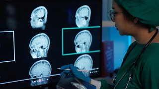
Brain signal processing: Searching for spikes
After the exam, Dr. Proskovec and I work with a team of physicians to analyze the (millions of) data points in the patient’s readout to look for tiny spikes in the wavy lines display. These spikes tell us where signals are misfiring in the patient’s brain and causing seizures. We approach signal processing like navigating with Google Maps. The head shape of 3D brain images is matched to your MRI to create the big picture of the map: the location, streets, and “neighborhoods” or sectors of the brain.
The MEG data is like the street-level information: How much traffic is there? How fast are the signals traveling? Is there a fender bender? Traffic jams and accidents register as spikes on the MEG data charts.
Running an algorithm against the data readout, we can locate the peak of the spike and match it down to the millimeter on the patient’s 3D brain map.
Then, we can use the maps to consult with the team of doctors about how they’ll approach the upcoming procedure.

How doctors use MEG data
Certain areas of the brain correspond to physical functions, such as motor skills, speaking, and sensory perception (like being able to perceive touch). Unfortunately, seizures can originate in or precariously close to these areas of the brain. In these cases, surgery can potentially alleviate the patient’s seizures, but altering these areas can result in long-term functional problems.
MEG-assisted mapping gives a brain surgeon more precise guidance than MRI and EEG alone can provide. EEG is useful to monitor brain activity, and MRI can show areas of potentially damaged tissue. MEG takes EEG to the next level by reporting much, much more activity and adding the spatial localization factor.
When combined with MRI, MEG can pinpoint the originating area of the seizure. Then we can consult with the team of doctors, including surgeons, neurologists, neuropsychologists, and radiologists, to determine a surgical plan that can relieve seizures, reduce functional risk, and allow the patient to maintain or improve their quality of life.
The limitless future of MEG technology
What we’re doing now at UT Southwestern with MEG imaging is so exciting. We have the first mixed use suite on campus – we do patient exams in the morning, and in the afternoon, we turn to research, pushing the boundaries of what the technology can do.
Future implementations of the technology are exciting, too.
Concussion research is huge in Texas, with our high volume of high school student-athletes. UT Southwestern is home to the ConTex Texas Sports Concussion Registry. In 2016, we conducted a concussion study using MEG to examine the brain function of high school football players. We used MEG to compare the network connectivity (how two or more brain regions “talk” to one another) of the brains of students who’d had at least one concussion during the season with those who hadn’t.
'We approach signal processing like navigating with Google Maps. The head shape of 3D brain images is matched to your MRI to create the big picture of the map: the location, streets, and “neighborhoods” or sectors of the brain. The MEG data is like the street-level information: How much traffic is there? How fast are the signals traveling? Is there a fender bender?'
Elizabeth Davenport, Ph.D.
We found that the concussed students had significant connectivity decreases, whereas the non-concussed students, on average, experienced increased connectivity. The results suggest that MEG could be more sensitive in detecting brain changes than functional MRI exams and should be researched further.
Alzheimer’s disease research is another area where we can potentially use MEG to identify biomarkers (brain or brain activity red flags) that can indicate a patient’s risk for mental health or cognitive conditions. I have a grant to study this. If we can discover a valid biomarker, doctors can potentially diagnose the condition and intervene earlier. Today, there is no cure for Alzheimer’s disease – we hope this research will pave the way for better treatments and potentially a cure.

Autism and PTSD are two other areas of study where we are using the MEG brain mapping technology to pursue discovery. In epilepsy and in future applications, MEG technology can help us advance diagnostics and research from “How can we find this variable?” to “Which of these millions of data combinations do you want to explore first?” That limitlessness is the challenge and the excitement of MEG technology.
Brain signal processing is an exploration of art and science. It drives us to push the boundaries of modern imaging and computing. And it keeps us asking, “What can we do next to improve the lives of patients with epilepsy?”
Are you or a loved one considering epilepsy surgery? Request an appointment to learn more about MEG brain mapping. Call 214-645-8300 or request an appointment online.