Magnetoencephalography (MEG)

Appointment New Patient Appointment or 214-645-8300

UT Southwestern Medical Center offers magnetoencephalography (MEG), a state-of-the-art technology that maps brain function. Our radiology specialists perform this testing before surgery to help plan procedures, particularly in patients with epilepsy.

UT Southwestern offers the most advanced MEG technology currently available, and we have the only MEG scanner in Dallas.

UT Southwestern Medical Center is recognized by U.S. News & World Report as one of the nation's top hospitals for neurological care.

Leading-Edge Technology Maps Brain Function

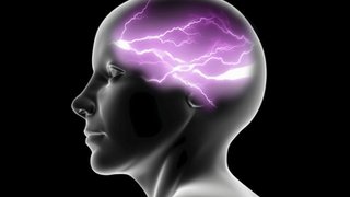
Magnetoencephalography (mag-ne-toe-en-sef-a-log-ruff-ee), also called a MEG scan or study, is a sophisticated, noninvasive brain imaging technology. A MEG scan is the newest, most advanced method for detecting, recording, and analyzing the magnetic fields produced by the brain’s electrical activity.

MEG studies are painless and safe for children and adults, requiring no needle injections, radioactivity, or strong magnetic fields. The MEG scan is completely silent.
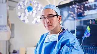
Our radiology experts at UT Southwestern are nationally recognized for their excellence in research and patient care. In our Neurodiagnostics Lab, we offer the latest innovations in diagnosis, using the most advanced technology available. We work to accurately diagnose epilepsy, brain tumors, and other brain disorders so that we can provide safe, effective treatment to improve our patients’ lives.

Magnetoencephalography: Why Is It Done?

At UT Southwestern, our brain specialists (neuroradiologists, neurologists, and neurosurgeons) use MEG scans to map brain function before brain surgery. We use MEG studies to identify the precise location in the brain where seizures or other problems arise.

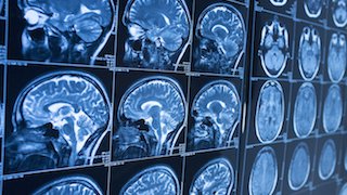
Magnetic resonance imaging (MRI) shows the brain’s structure, and we combine MRI with MEG. The combined images map the areas of normal and abnormal brain activity. We use MEG scans to make brain surgery safer and more effective by targeting areas for treatment and avoiding surrounding healthy tissues.

Our neuroradiology specialists use MEG scans to identify and map:

  • Areas of the brain that control language, memory, motor, and sensory activities
  • Areas of the brain where epileptic seizures begin
  • Specific locations of brain tumors and other lesions

We often recommend MEG studies for people who are undergoing brain surgery for conditions such as:

To schedule a MEG at UTSW please reach out to our outpatient imaging team: 214-645-7237.

What to Expect

We’ve provided these recommendations to help patients prepare for the MEG scan. Please contact our care team at 214-645-0681 or email if you have any questions.

Please notify us if you or your child has:

  • An electronic implant, such as a pacemaker or a vagus nerve stimulator (VNS)
  • Metal braces
  • A permanent retainer
  • Permanent makeup or lash extensions
  • Lice and nits are visible in the hair (we will reschedule the study)

How to prepare:

  • Please complete the screening form in MyChart, and we will review it with you when you arrive.
  • For epilepsy focus mapping: Please decrease sleep the night before the test. The patient must be ready to sleep during the scan. The results are more successful if the patient sleeps during the study.
  • Patients can wash their hair the night before the test but should leave their hair loose and not use any hair products.
  • Patients can wear contact lenses but not glasses during the scan.
  • Eat breakfast.
  • Stay on your medication regimen.

Please bring the following items:

  • Any rescue medications
  • List of current medications
  • Disc of brain MRI, if not completed at UT Southwestern, Parkland, or Children’s hospitals
  • If the patient wears glasses, we will need the glasses prescription
  • Snacks
  • Driver’s license, insurance card, and form of payment
  • Children can bring a comfort item, such as a bottle, pacifier, toy, or blanket, to hold during the test. The item must not contain metal.

Please avoid these items:

  • Caffeine or candy after midnight of the night before the test or the morning of the test
  • Hair products, hair extensions, and hair weaves
  • Makeup, lotions, or other skin-care products
  • Metallic fingernail polish
Checking in for the procedure

On the day of the appointment, please:

  • Arrive 15 minutes before the appointment.
  • Check in with the Radiology Department on the second floor of the Clements Imaging Building (Rogers MRI).
After check-in

Once you check in and before the patient enters the MEG scanner:

  • We will ask the patient to change into hospital scrubs.
  • A locker will be provided to secure any belongings.
  • No metal can be taken into the MEG scanner. This includes:

    • Electronic items such as games, movies, or music while in the MEG scanner
    • Jewelry or watches
    • Clothing or undergarments that have metal, such as zippers, underwire, or snaps
During the procedure

To begin the procedure:

  • Our care team puts paste or gel in the patient’s hair and places an EEG cap on the patient’s head.
  • The care team will then touch the cap and the patient’s nose and ears with a wand to help the computer create a picture of the head.
  • We place the patient in the MEG, a scanner with hundreds of specialized, magnetic sensors around, but not touching, the patient’s head. The MEG sends signals to a computer in another room.

During the MEG scan:

  • The patient will sit or lie on a table in the MEG scanner room.
  • Parents or others accompanying the patient can wait in the radiology waiting room. If your child needs a family member during the procedure, we can make special arrangements.
  • The technologist will be in constant communication with the patient while the study takes place.
  • We typically record brain activity in both adults and children while awake and asleep. Depending on the reason for the test, we might ask the patient to sit still, lie down, or complete various simple tasks during the wakeful portion. 
After the procedure

The exam is an outpatient procedure that typically takes about one to four hours, based on the test your physician orders. Patients can leave shortly after the exam and resume their normal activities.
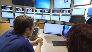
Our neuroradiology and neurology team will work together to review the MEG recordings. The physician who ordered the MEG scan will receive the results.

Research

UT Southwestern’s physician-scientists use MEG technology in ongoing research to improve patient care, with studies that help:

  • Locate, before surgery, the sources of epileptic seizures to improve surgical outcomes of patients.
  • Locate, before surgery, the eloquent cortex in patients undergoing brain surgery for trauma or epilepsy. The eloquent cortex refers to areas of the brain that, if removed, would result in the loss of motor skills, sensory processing, linguistic ability, or paralysis.
  • Diagnose concussions and traumatic brain injuries (TBIs), which cannot be diagnosed with MRI.
  • Map brain networks to study PTSD and other brain disorders.
  • Conduct research on the effectiveness of medications to treat schizophrenia, a mental disorder that affects thought, emotion, and behavior.
  • Diagnose Alzheimer’s disease before symptoms appear.
  • Diagnose autism and better understand processing in the brains of people with autism.

Referring Providers

If you are a referring physician, please use this form to place an order.