How bone optimization, novel procedure help heal pelvic insufficiency fractures
September 24, 2024


When you picture a broken pelvis, you likely envision a severe fracture caused by a high-impact accident, such as a vehicle crash or falling from a ladder. These fractures are easy to identify – they appear as large breaks on X-rays.
But in patients age 50 and older, particularly women, it doesn’t take a considerable trauma to fracture the sacrum, a triangular bone structure that connects the spine to the pelvis.
These pelvic insufficiency fractures can happen after a low-impact accident such as falling from a standing height if a patient’s bones are weakened by osteoporosis, vitamin D deficiency, rheumatoid arthritis, or pelvic radiation therapy. These insidious breaks, classified as insufficiency fractures, can linger undetected for months because they aren’t as keenly visible in imaging.
While pelvic insufficiency fractures can heal on their own over time, they cause persistent pain in the low back, groin, and buttocks, as well as decreased mobility. Often, they heal as “nonunion” fractures in which the bones don’t rejoin properly, inviting future complications. Pelvic insufficiency fractures are also linked to increased mortality – research has shown a one-year mortality rate of 23% post-fracture in people 65 and older.
Treatment for pelvic insufficiency fractures used to be limited to pain medication and physical therapy, if the break was recognized at all. But today, an innovative procedure – paired with advanced bone optimization therapy – can significantly relieve pain during healing and reduce the risk of future fractures.
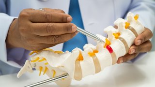
Percutaneous sacral fixation is a fast, streamlined procedure that involves rejoining the fractured pelvis with a screw-and-nut orthopedic system, with no need for large incisions or open surgery.
UT Southwestern is a pioneer of this procedure and has launched a unique program that bookends surgery with endocrine and bone optimization therapies through our Mineral Metabolism program. By strengthening the bones before and after surgery, we can offer patients lasting relief and strategies to potentially avoid painful fractures in the future.
It is our priority to make more North Texas health care providers aware of this innovative approach. Over 10 million Americans have osteoporosis, and half of women and a quarter of men over age 50 will break a bone due to this condition in their lifetimes. We expect these numbers to increase as the population ages. Our hope is that spreading the word will help more patients access this pain-relieving treatment program.
How are pelvic insufficiency fractures diagnosed?
An initial physical exam will indicate pain at the site of the fracture. Other symptoms might include:
- Difficulty urinating.
- Pain in the abdominal area.
- Tingling or numbness in the leg or groin.
In addition, patients with a pelvic insufficiency fracture often cannot complete the Timed Up and Go test (TUG). In this test, patients are timed getting up from a seated position, walking 10 feet, walking back, and sitting down. If the total test takes more than 30 seconds or they cannot rise, the patient likely has a mobility complication.
While a pelvic insufficiency fracture is not easily visible with an X-ray, CT scan and MRI scan are highly sensitive and should be considered in patients with pelvic pain who are unable to mobilize.
If a pelvic insufficiency fracture is confirmed, your provider will likely screen you for osteoporosis and other bone disorders using a DXA scan, low-dose X-ray technology to measure bone density. DXA scans can tell providers if a pelvic insufficiency fracture is part of a larger pattern of bone fragility – the lower the density, the greater the risk of fractures.
"Our goal is to raise awareness so more patients can find pain relief sooner – and optimize their bone health to proactively reduce the risk of pelvic insufficiency fractures."
Ishvinder Grewal, M.D.
How does percutaneous sacral fixation work?
Percutaneous sacral fixation is a minimally invasive surgery that involves two to four incisions the size of a thumbnail where the low back meets the pelvis. A screw is inserted to hold the fractured bone together and a nut applied on the far side secures it, compressing the sacrum to prevent shifting and decrease pain during healing.
For younger patients with high-energy fractures, we sometimes remove the screws after healing. For older patients, we often keep the screws in place to provide added long-term stability.
For many patients, this procedure can reduce pain and accelerate recovery time. It should be considered instead sacroplasty, in which bone cement is injected to fill a sacral fracture. Sacroplasty is not recommended by pelvic surgeons for patients with sacral fractures, since it does not provide mechanical stability, prevents fracture healing, and can obscure pathways for screw fixation. Additionally, cement cannot be removed from the sacrum once injected. In fact, Milton Lee Routt, Jr., M.D., one of the most eminent pelvic trauma surgeons in the world, took to LinkedIn in September 2024 to urge doctors not to practice sacroplasty in these patients.
Percutaneous sacral fixation is currently offered only at advanced orthopedic centers like UT Southwestern. We currently perform 50 to 100 procedures a year. Our goal is to raise awareness so more patients can find pain relief sooner – and optimize their bone health to proactively reduce the risk of pelvic insufficiency fractures.

Why does bone mineral optimization improve outcomes?
While subcutaneous sacral fixation can reduce pain, it doesn’t improve overall bone health. But specialists in UTSW’s Mineral Metabolism program can help patients build a solid foundation for the procedure that supports healing while also reducing the risk of future pelvic insufficiency fractures.
Each patient gets a customized treatment plan based on our three-pronged approach to bone optimization.
1. Nutrition and mineral support
Patients can work with providers in our Mineral Bone Health program to incorporate these bone-strengthening vitamins and minerals into their diet:
- Calcium makes up the bone matrix, a major component of overall bone structure. Calcium deficiencies can cause weaker, more porous bones.
- Vitamin D helps the digestive system absorb calcium.
- Phosphorus combines with calcium to give bones their strength and rigidity. High phosphorus intake can reduce the risk of osteoporosis by 45%.
- Magnesium helps regulate calcium and vitamin D levels, preventing deficiency or excess buildup.
Patients 65 and older are automatically enrolled in RESTORE – Returning Seniors to Orthopedic Excellence, which focuses on continued care after a fragility fracture. RESTORE follows patients from admission through six to eight weeks after discharge, bringing together experts in pain management, orthopedics, and geriatrics to maximize function and reduce the risk of future fractures.
2. Physical therapy and exercise
Patients can work with a physical therapist or primary care provider to create an exercise plan that reduces the risk of fracture while strengthening bone. Movement as a treatment for osteoporosis may seem counterintuitive since intense physical activity can cause osteoporotic fractures in patients with low bone density. However, resistance training and weight-bearing exercises can improve overall bone health through mechanical stress, which signals the body to start building new bone tissue.
One study found that brisk walking more than four hours a week may reduce the chance of hip fractures by 41%, while another discovered that lifting weights and high-impact exercise can improve bone mineral density up to 4% per year in women. Gentle movement, such as yoga or water aerobics, can improve balance and coordination to decrease the chance of falls.
3. Hormone therapy
Supplemental estrogen therapy can help slow bone loss during perimenopause or menopause, when the risk of osteoporosis increases. High levels of thyroid hormones can lead to osteoporosis by increasing the breakdown of bone faster than new bone can form. Some of the treatments that can regulate thyroid levels include:
- Anti-thyroid medications such as methimazole, carbimazole, or propylthiouracil, which block a key enzyme that thyroid hormones need to replicate.
- Radioiodine treatment, which is absorbed by the thyroid gland to destroy some of the thyroid tissue and slow hormone production.
As the population ages, we will see the rate of pelvic insufficiency fractures increase. Our goal is to inform patients and providers in the community to be aware of these dangerous fractures – to know how to avoid them, how to diagnose them, and how this minimally invasive surgery and bone health program can reduce the risk of future fragility fractures.
To talk with a specialist about pelvic insufficiency fracture care, call 214-645-8300 or request an appointment online.