Seeing the invisible: How we use advanced technology in eye exams
January 24, 2019


Doctors generally aren’t satisfied with the status quo. It’s part of our nature. We push for the latest advancements, the newest treatments, and the most cutting-edge options available to continually improve the care we’re able to provide to our patients.
Neither of us is an exception to this, nor are our colleagues in the Department of Ophthalmology at UT Southwestern. For more than four decades, our department has been recognized as a national and international leader in caring for patients with the most challenging conditions and diseases that affect the eye. Both of us specialize in vitreoretinal diseases, which are disorders that affect the retina (the part of the eye that receives images and transmits them to the brain through the optic nerve) and the vitreous (the clear gel that lies between the front of the eye and the retina).
Because of our expertise, many other eye doctors refer patients with unusual or complex conditions affecting the retina or vitreous to us. One reason we’re ready and able to care for these patients is our dedication to incorporating the latest technology into our diagnostic efforts. We also regularly participate in nation-wide clinical trials for new potential treatments.
Optical coherence tomography: A major advancement
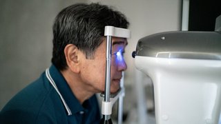
Optical coherence tomography (OCT) is probably the biggest diagnostic breakthrough in ophthalmology in the past 20 years. This noninvasive test allows us to see each of the individual layers of the retina, so we can determine any anatomic abnormalities. The advanced OCT machines we currently use provide almost as much detail as would be obtained by complex histological studies… histological studies are clearly not an option in clinical care since they would involve removing the eye.
OCT has revolutionized how we diagnose and treat our patients. We previously had to consider some patients’ conditions as unexplained because we couldn’t see enough of the details of how their eyes were affected. With OCT, we can gather huge amounts of detail, which aids us in the diagnosis and treatment of many conditions, such as:
● Diabetic retinopathy, a complication of diabetes that involves damage to the retina’s blood vessels
● Macular degeneration, typically an age-related degeneration that causes damage to the central retina that results in loss of central vision
● Vitreomacular traction, caused by pulling on the retina by the vitreous that can blur or distort vision
We are fortunate to have three distinct OCT imaging devices available to us at UT Southwestern. Each machine is linked to different databases and lends itself to different specialized usages for our patients. For example, one of our OCT devices is dedicated to OCT angiography, which lets us see the network of blood vessels that bring blood to the retina and optic nerve. This is extremely useful for diagnosing and treating retinal diseases, particularly those caused by macular degeneration and diabetes. One of our newest OCT machines is based on swept-source technology, which uses a specialized, tunable laser that allows us to better visualize the vitreous. This enables us to better understand and diagnose vitreomacular traction and other diseases that affect the vitreous.
"We push for the latest advancements, the newest treatments, and the most cutting-edge options available to continually improve the care we’re able to provide to our patients."
– Zachary Robertson, M.D., and Rafael Ufret-Vincenty, M.D.
Electrophysiology testing for the eye
Patients most often hear about electrophysiology as it applies to the heart’s electrical system. However, ophthalmic electrophysiology, also known as visual electrophysiology, deals with the eye’s electrical activity. When light enters the eye, the retina converts the image of what you see into an electrical signal that it transmits to the optic nerve, which carries the electrical signal to the brain.
Electrophysiology tests allow us to assess how well this process works. So, while OCT gives us anatomical information about the retina (how it looks), electrophysiology gives us functional information (how it works). To do this, our team uses different versions of a test called an electroretinogram, or ERG. A full-field ERG measures the electrical response of the entire retina at once, while a multifocal ERG measures responses from many different areas of the retina at the same time for pinpoint accuracy of where a patient might be experiencing retinal issues. These tests often aren’t available at smaller, private ophthalmology practices, but they can be key to helping us understand conditions that we can’t see with our own eyes or through other imaging tests.
ERGs are also helpful for monitoring patients on high-risk medications, such as hydroxychloroquine. This medication can damage the retina and lead to loss of vision, so we have to check the retinas of patients on hydroxychloroquine to make sure their retinas are healthy. Doctors might prescribe hydroxychloroquine to treat the following conditions:
- Lupus, a disorder of the immune system that involves inflammation of various areas of the body
- Rheumatoid arthritis, an inflammatory disorder that primarily affects joints
- Sjögren’s syndrome, a disorder of the immune system characterized by dry eyes and dry mouth
Artificial intelligence: The future of retinal ophthalmology?
In April 2018, the Food and Drug Administration (FDA) approved IDx-DR, a software program that uses an advanced artificial intelligence algorithm to analyze images of patients’ retinas for signs of diabetic retinopathy after a doctor loads them into the program. If IDx-DR determines that a patient is at high risk for the condition, it instructs the doctor to refer the patient to an eye-care professional for further screening. According to the FDA’s analysis of the program, IDx-DR was able to correctly identify diabetic retinopathy nearly 88 percent of the time, and it was able to successfully rule out the condition nearly 90 percent of the time.
Of course, we are a long way from having robot ophthalmologists assisting us in the exam room. But we think ophthalmology will embrace revolutionary ideas like IDx-DR in the years to come. Programs and devices with deep-learning capabilities could potentially notice and quantify data points that are still invisible to doctors’ eyes. In the future, this might be able to assist us in helping more people at earlier stages of eye disease.
The future is never far away for us in ophthalmology. In fact, we’re constantly pushing toward it by testing the newest innovations and working to incorporate the latest technology into our advanced care in ways that will most benefit the patients we serve.
To find out whether you or a loved one might benefit from retinal ophthalmology screening, call 214-645-2020 or request an appointment online.
Related Clinics
Ophthalmology
at UT Southwestern Monty and Tex Moncrief Medical Center at Fort Worth 600 South Main Street, 1st Floor, Suite 1.500Fort Worth, Texas 76104 817-429-3050 Directions to Ophthalmology at UT Southwestern Monty and Tex Moncrief Medical Center at Fort Worth, Fort Worth Parking Info for Ophthalmology