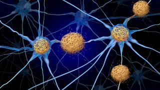
The brain is a complex, sophisticated network – the origination point for every physical and mental task we perform. Seemingly simple actions such as giving a thumbs up or saying hello are driven by neurons firing in the brain and sending signals throughout the body.
So, when part of that network is damaged or disrupted, the result can be conditions such as epilepsy, Parkinson’s disease, and severe depression, which compromise our ability to move, think, or process emotions.
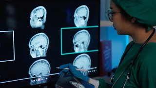
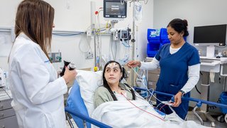
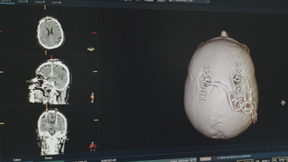
But leaps forward in brain mapping technology are making it possible to better understand the structure and functions of the brain. Neurosurgeons can place tissue-stimulating electrodes precisely and significantly relieve symptoms such as seizures and tremors.
During my career, I have helped advance brain mapping techniques to personalize our approach to neurological care. One of my most well-known patients was an actor and musician whose essential tremor had significantly reduced his ability to play guitar – and he was concerned that brain surgery would rob him of it completely.
So, we invited him to strum his guitar while we performed a brain mapping procedure called deep brain stimulation to precisely target and treat the source of his symptoms. The surgical team was excited to provide a more personalized treatment, and the patient was thrilled to maintain his ability to make music.
At UT Southwestern, our neurosurgery department is at the forefront of brain mapping research and clinical implementations. One of our team’s greatest strengths is understanding the root cause of an illness and finding the right treatment to improve function for our patients.
Brain mapping and advanced imaging play a key role by giving us the ability to:
- Safely investigate the inner workings of the brain
- Plan precise brain surgeries and improve therapies to relieve patients’ symptoms
- Measure the effectiveness of treatment using biomarkers – physical cues that are personalized to each patient’s brain
Where brain maps lead
UT Southwestern offers a vast repertoire of tools to study and map the brain. Some of those include:
- Intracranial electroencephalography (iEEG): By placing electrodes strategically inside or in contact with the surface of the brain, we can record its electrical activity to learn precisely where network disruptions are occurring and what symptoms they cause, such as tremors or depressive episodes.
- Intraoperative electromyography (EMG): Thin needles are inserted into the muscles to record electrical activity. This information tells us whether stimulation in certain areas of the brain affects muscle function.

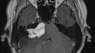
- Magnetoencephalography (MEG): This sophisticated technology detects, records, and analyzes magnetic fields produced by the brain’s electrical activity. Through an external helmet and with no insertion of electrodes into the brain, MEG can show us specifically where seizures originate, where lesions or tumors exist, and which tissues we should avoid to prevent loss of motor, memory, or language functions.
- Nerve conduction studies: Small electrical shocks stimulate the nerves, and we record the signal that those nerves produce. This information can show whether electrical signals are traveling along the nerve as expected.
- Diffusion tractography: This advanced MRI technology shows us white matter pathways (communication channels) and how they are anatomically associated with diseased and damaged brain tissue. The imaging works by identifying where water molecules spread out in the brain – they spread more easily through the channels than across them.
- Functional magnetic resonance imaging (fMRI): We use noninvasive fMRI to detect changes in blood flow as a measure of brain activity. When blood flow is high, the area of the brain shows brighter on the fMRI due to a higher volume of blood flow.
Related reading: MEG technology: Improving epilepsy surgery outcomes, one weird helmet at a time
Planning and improving therapies
Using the information gained from brain mapping, we can plan and personalize treatments for conditions that don’t respond to standard therapies. Some of these technologies include:
Deep brain stimulation (DBS): This procedure involves permanently placing electrodes in the brain that can be activated either automatically or manually by an external unit to stimulate specific areas of the brain that are the “cogs” in the neural communication network. DBS can be used independently or in combination with technologies such as high frequency ultrasound (HIFU) to remove damaged tissue.
DBS is an approved therapy for certain conditions, including focal epilepsy (seizures that affect one side of the brain), Parkinson’s disease, essential tremor, and dystonia. Through advanced brain mapping such as fMRI, we can more precisely place the electrodes, further personalizing an already effective treatment to significantly reduce tremors and seizures.
We are actively expanding DBS as a treatment option for network-based conditions with less visible symptoms, such as severe depression. By pairing DBS with advanced imaging, we can map areas of a patient’s brain to understand where network disruption occurs and stimulate those regions to realign the brain’s electrical rhythms – much like a pacemaker or defibrillator restores a natural hearth rhythm.
Electroconvulsive therapy (ECT): With this therapeutic procedure, a patient is placed under general anesthesia. The neurologist induces 30- to 45-second medically controlled seizures using electrical currents for a total of about five minutes. The seizures are thought to affect specific neurotransmitters or circuits in the brain associated with mental health symptoms.
ECT is approved by the U.S. Food and Drug Administration (FDA) to relieve treatment-resistant depression. In patients who are good candidates, it is 80% effective and can provide full reversal of depression symptoms in some patients. ECT can also treat psychosis and catatonia, a condition that interferes with speech and movement.
Transcranial magnetic stimulation (TMS): This evidence-based treatment for severe depression uses a metal coil placed below the scalp to deliver gentle magnetic pulses to the brain in hopes of altering the function of nerve cells in the brain, improving and potentially resolving severe depression symptoms. Treatments are delivered in approximately 30 sessions over six weeks, each lasting 15 to 30 minutes.
Related reading: Using magnets to treat major depressive disorder - clinical trial shows promise
Brain Power
Advances in brain mapping, CRISPR technology, and neurosurgery are being transformed into life-changing treatments for patients at UT Southwestern’s Peter O’Donnell Jr. Brain Institute. Dr. William Dauer, Director of the Institute, and Dr. Nader Pouratian, Chair of Neurological Surgery, discuss how their teams are unlocking the mysteries of brain diseases, from epilepsy to Alzheimer’s.
Identifying biomarkers to measure treatment
For conditions with tangible symptoms such as tremors, seizures, and the inability to control muscle movement, treatment success if obvious. The patient’s tremors are significantly reduced, and they can better control their movements.
For conditions with less visible symptoms, such as depression, there are limited outward markers of success. We must rely on a patient’s self-reported symptom improvement and their ability to participate in daily activities. And while they may start feeling better, it’s difficult to know if more improvement is possible.
However, for certain conditions, advanced brain mapping allows us to identify physical biomarkers – areas or communication channels in the brain that we expect to change with treatment – to measure therapy effectiveness and guide future treatments.
The future of advanced brain mapping
Brain mapping technology continues to evolve rapidly, increasing our ability to make a marked difference in more patients’ lives.
One area of innovation combines advanced endovascular techniques with brain mapping technology. It may be possible one day to place electrodes through a patient’s blood vessels rather than their brain tissue, allowing us to monitor their brain through the electrical activity in the blood vessels. Though such a procedure would be delicate and there would be a risk of clotting, endovascular electrode placement could become a less-invasive option to manage complex brain conditions.
At UT Southwestern, we consistently host and participate in clinical trials that can help advance the field of brain mapping. Currently, we are participating in two clinical trials for DBS for depression, and we are planning to launch another for DBS to treat facial pain.
We also continue to refine and discover treatments for epilepsy, movement disorders, mental health conditions, and other network-based conditions.
The goal for treatment is to improve brain function and help patients enjoy more days where they can brush their own teeth, have the energy to get out of bed, and simply feel comfortable in their own bodies. They may still have seizures, tremors, or depression episodes, but we are working to make their symptoms less frequent and severe.
Through advanced brain mapping techniques, we can restore some sense of normalcy and predictability to the lives of our patients.
To talk with a neurosurgeon about advanced brain mapping, call 214-645-8300 or request an appointment online.