The ‘rare’ Porzingis ankle injury: What is it and how is it treated?
June 14, 2024


When the spotlight is the brightest and the stakes are the highest in professional sports, any injury to a star player gets magnified.
Cases in point: In the 2023 NFL playoffs, Kansas City Chiefs quarterback Patrick Mahomes suffered a high ankle sprain during the divisional round and the sports world held its breath until he hobbled back onto the field and eventually led his team to the Super Bowl. The next season, New York Jets quarterback Aaron Rodgers was sacked early in the first Monday night game, suffering a season-ending torn Achilles tendon.
Fast forward to the 2024 NBA Finals, when Boston Celtics 7’2” center Kristaps Porzingis suffered an injury to his size-16 left foot that is nearly as uncommon as the mythical creature for which he’s nicknamed, “The Unicorn."
Porzingis sat out for Games 3 and 4 against the Dallas Mavericks, his former team, due to a “torn medial retinaculum, allowing dislocation of the posterior tibialis tendon” (PTT), according to a team statement.
Porzingis logged some playing time in Game 5, where the Celtics outscored the Mavericks to secure their 18th NBA title. On June 27, the team put out a statement, saying Porzingis "underwent successful surgery to repair a torn retinaculum and dislocated posterior tibialis tendon" and is expected to be out for 5 to 6 months.
The medial (flexor) retinaculum is a 1 millimeter-thick strip of connective tissue that holds the arch of the foot in place like a sling and allows for jumping, running, and raising on the toes. The injury threatens the versatile footwork, quick cutting, and on-court agility that earned Porzingis his “unicorn” status and made the Latvian baller a first-round 2015 NBA draft pick.
While sprains and tears of the lateral ankle ligaments on the outer edge of the ankle are commonplace, medial ankle injuries are rare. Injuries of the medial retinaculum are even less common when compared to deltoid ligament injuries – I've seen just one in my career that involved a dislocated PTT. It’s rare because the overextension occurs on the inside of the foot, which is an awkward movement in an area that is typically stout. Medial retinaculum soft tissue damage is usually associated with high-energy trauma, such as a car accident or falling during a fast movement.
Though uncommon, a torn flexor retinaculum can be healed with proper treatment ... and patience.
What’s going on in the foot?
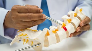
Medial retinaculum tissue supports the fingerlike long bones of the foot and anchors the 7 centimeter-thick posterior tibialis tendon (PTT) – the largest of the flexor tendons in the ankle – from the medial malleolus (the inner knobby bone of the ankle) onto the calcaneus (heel bone).
A tear in the retinaculum from a traumatic foot or ankle injury lifts the tissue from the ankle bone or strips it entirely. This weakens the sling, causing pain in the inside edge and back of the ankle and foot, along with instability, especially while weight-bearing or doing power moves like jumping and running.
Medial retinaculum tears increase the risk that the PTT will slide out of place, changing the trajectory of an ankle in motion and limiting the ability to flex the foot or walk. While traumatic dislocation of the PTT is rare, it is the second-most common type of ankle tendon dislocation.
A medial retinaculum tear can easily be misdiagnosed as an ankle sprain. When the ankle is swollen, it’s difficult to clinically evaluate for the injury. But after the swelling goes down, patients may experience telltale signs such as ongoing pain or swelling with activity, a “snapping” sensation over the bone on the inside edge of the ankle, or an increasingly flattened arch over time.
Treatment options for flexor retinaculum tears
Nonoperative treatment
The good news is nearly all medial retinaculum tears can heal without surgery. Treatment for a pro athlete like Porzingis will be expedited, but for the rest of us, a slow-and-steady approach over three to six months is the best path to full recovery.
Once the swelling goes down in the ankle, usually about one to two weeks after injury, we conduct a physical exam to assess where it hurts and how much. If indicated, an MRI of the ankle may then be performed to identify the extent of tissue damage.
In patients with low grade tears and stable tendons, they will spend four to six weeks in a boot to immobilize the tissue as it heals, and we recommend the RICE method – rest, ice, compression, and elevation – during this time to optimize healing.
Next, we move to a brace and begin physical therapy. The UT Southwestern Sports Medicine and Physical Medicine and Rehabilitation teams work with professional, collegiate, and everyday athletes to start improving agility and stability early in a foot or ankle injury, reducing long-term instability and mobility limitations. Within three months, patients can ease back into impact exercises, and by six months, most athletes can safely return to regular play.
Surgical treatment
Surgery may be appropriate in severe medial retinacular tears or in cases where nonoperative therapies do not provide sufficient stability and pain relief. The UTSW Orthopaedic Surgery department offers the latest technology and techniques, including minimally invasive surgery to repair a torn medial retinaculum by reattaching it to the ankle bone or “battening down” tissue that is still attached but lifted from the bone.
The gap that forms between loose tissue and bone is called a hematoma, which is essentially a pouch where the PTT can slip into from its normal position behind the medial malleolus.
If the PTT has slipped already, we can reposition it. In rare cases, we may need to create a deeper groove in the ankle bone and suture the retinaculum to prevent future displacement. After this outpatient procedure, patients will spend about six weeks in a boot or splint. Over time, the tendons and retinaculum tissue will loosen up, so we make a tight sling to support longest-lasting stability.
Surgery does not heal the injury – it merely puts things back where they are supposed to be. So, a diligent physical therapy plan is essential for proper, timely recovery. After surgery, we see patients every two to three weeks to help them stay on track for recovery.
Related reading: High ankle sprain vs. low ankle sprain: What are the key differences?
Getting back in the game safely
Once you have a soft tissue injury in the ankle, you are more susceptible to having another one. When you return to activity, we encourage you to reduce your risk of further sprains, strains, and tears by:
- Stretching before playing: Talk with your doctor and physical therapist about proper warmup movements for your sport and anatomy.
- Wearing proper gear: Shoes with ample support, custom orthotics, or a brace can provide stability while you exercise.
- Paying attention to your body: If you hear or feel a snapping sensation across the inner bone of your ankle or pain in the inner or back of the joint, talk with your doctor to avoid further tissue damage.
If you follow your treatment and rehab plan, you should be back on the court, field, or track before you know it – with a strong, stable ankle.
To talk with an orthopedic specialist or sports medicine provider, call 214-645-8300 or request an appointment online.