

Find a Clinical Trial
Search for opportunities to participate in a lung disease or asthma research study.
New Patient Appointment or 214-645-7700
The thoracic surgeons and interventional pulmonologists at UT Southwestern Medical Center use leading-edge methods to evaluate and treat pulmonary nodules and other various lung lesions – including bronchoscopic procedures, image-guided sampling, conventional surgery, and more advanced minimally invasive and robotic techniques.
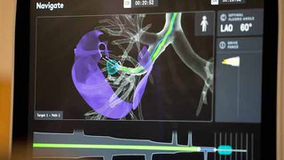
We feature the latest imaging techniques and treatments through advanced imaging, including endobronchial ultrasound (EBUS), robotic bronchoscopy, and many others.
Lung nodules and lung lesions are spots or growths on the lungs.
Lung nodules can be benign (noncancerous) or malignant (cancerous). Lung nodules rarely have symptoms and are often incidentally found. They can represent an early opportunity to identify and treat lung cancer.
The most common causes of benign nodules include granulomas (clumps of inflamed tissue) and hamartomas (benign lung tumors).
The most common cause of cancerous or malignant lung nodules includes lung cancer or cancer from other regions of the body that has spread to the lungs (metastatic cancer).
Up to half of those diagnosed with early-stage lung cancer are identified through an incidentally detected lung nodule. However, not all incidental lung nodules are cancerous.
Where patients go first for diagnosis and treatment truly matters. Our surgeons work closely with UT Southwestern’s interventional pulmonary team, oncologists, radiologists, and pathologists to deliver comprehensive care – all in one location.
From the time of first referral, patients can expect to see a provider within a week. The lung nodule clinic at UT Southwestern streamlines and expedites care to identify the correct treatment for patients.
The risk of a lung nodule being cancerous varies considerably depending on several things, including:
If we suspect a patient has a lung nodule, we will conduct a physical examination and order tests to confirm the diagnosis. Studies to evaluate and diagnose pulmonary nodules might include:
Based on the characteristics and size of the lung nodule on the CT scan, we may recommend:
When surgery is the most appropriate therapy, our thoracic surgeons treat lung nodules and lung lesions with procedures that include:
Compared with surgery performed through an open chest incision, minimally invasive surgery provides several important benefits for patients, including:
When surgery is not possible in a patient with a cancerous or malignant nodule, our multidisciplinary team of surgeons and medical and radiation oncologists will provide recommendations about the best management options, which may include advanced radiation techniques, systemic therapy with conventional chemotherapy, and/or targeted or personalized therapies.
UT Southwestern conducts clinical trials aimed at improving the treatment of lung nodules. Talk with our doctors to see if a clinical trial may be available.
Search for opportunities to participate in a lung disease or asthma research study.
We’re one of the world’s top academic medical centers, with a unique legacy of innovation in patient care and scientific discovery.