Search for opportunities to participate in a clinical research study.
Ultrasound
New Patient Appointment or 214-645-8300
UT Southwestern Medical Center’s ultrasound services allow physicians and specialists to make accurate diagnoses and prescribe targeted treatments and therapies for a wide range of conditions, from the simple to the complex.
Our radiology team performs more than 800,000 inpatient and outpatient exams every year. We specialize in advanced technologies and the latest clinical innovations in today’s changing field of medical imaging
Fast, Safe, and Reliable Imaging
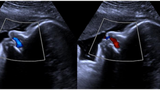
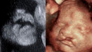
Ultrasound, or sonography, is a scan that uses high-frequency sound waves to show the inside of the body. The ultrasound reveals movement and live function in the body’s organs in real time. The test is safe and easy and does not use X-rays or radiation.
Because the body is more than 90 percent water, sound waves can travel through it just as sonar is used in the ocean. As sound waves from the ultrasound machine go through the body, they create an echo when they hit various tissues. The returning echoes are recorded by a computer that then displays them on a screen to an ultrasound technologist.
UT Southwestern offers experience and expertise in all types of ultrasound, including technologies and techniques that might not be available at other medical facilities.
Conditions We Diagnose with Ultrasound
We can use the results of an ultrasound to detect and diagnose a wide range of medical conditions.
Ultrasound is most effective in diagnosing conditions by:
- Examining the heart
- Evaluating vascular disease
- Revealing information about the size and shape of tumors and cysts
- Evaluating the gallbladder and related organs
- Evaluating the uterus and ovaries
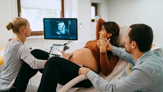
- Examining the fetus during pregnancy
Ultrasound: What to Expect
An ultrasound is safe and painless. Patients might be asked to fast for several hours before the exam or drink several glasses of water to create fullness in the bladder. We will give patients any specific instructions they need.
Patients should avoid carbonated beverages before the exam, because bubbles in the body can interfere with the ultrasound images. An ultrasound technologist can answer any questions a patient might have about a health condition that could affect the exam.
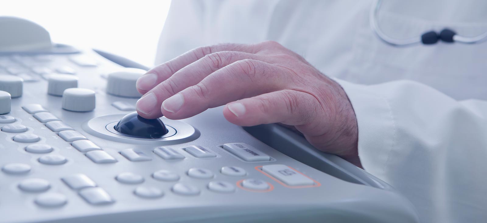
An ultrasound exam usually takes 30 minutes. The technologist will ask the patient to lie or sit on an examination table. The technologist can then lower the lights in the room to make the computer display easier to see. A gel will be applied to the patient’s skin over the area to be scanned. This gel allows the ultrasound transducer, which transmits images to the computer, to slide easily over the skin.
The patient might feel some discomfort if he or she has a full bladder and the technologist is pressing the transducer wand over the abdomen.
For pelvic examinations, such as those for the prostate gland, uterus, or ovaries, the technologist will explain the use of an ultrasound probe. This probe is placed in the rectum or vagina to better capture images of internal structures. Patients can ask for a third person, or chaperone, to be present at these types of intimate exams, if they wish.
As the transducer transmits live images of the patient’s body to the computer, the technologist will capture pictures for permanent reference.
A radiologist will review the images and send a report to the doctor, who will notify the patient of any findings. The patient can also request to receive the images on CD.


We’re one of the world’s top academic medical centers, with a unique legacy of innovation in patient care and scientific discovery.

MedBlog


Results: 7 Locations
Cardiology
at UT Southwestern Medical Center at Park Cities 8611 Hillcrest Road, 2nd FloorDallas, Texas 75225 214-692-3135 Directions to Cardiology at UT Southwestern Medical Center at Park Cities, Dallas Parking Info for Cardiology
Professional Office Building 2
5939 Harry Hines Blvd.Dallas, Texas 75390 214-645-8300 Directions to Professional Office Building 2, Dallas Parking Info for Professional Office Building 2
Clinical Heart and Vascular Center
at West Campus Building 3 2001 Inwood Road, 5th FloorDallas, Texas 75390 214-645-8000 Directions to Clinical Heart and Vascular Center at West Campus Building 3, Dallas Parking Info for Clinical Heart and Vascular Center
Imaging Services
at UT Southwestern Medical Center at RedBird 3450 W. Camp Wisdom RoadDallas, Texas 75237 (214) 645-9729 Directions to Imaging Services at UT Southwestern Medical Center at RedBird, Dallas Parking Info for Imaging Services
Ophthalmology
at UT Southwestern Monty and Tex Moncrief Medical Center at Fort Worth 600 South Main Street, 1st Floor, Suite 1.500Fort Worth, Texas 76104 817-429-3050 Directions to Ophthalmology at UT Southwestern Monty and Tex Moncrief Medical Center at Fort Worth, Fort Worth Parking Info for Ophthalmology
Ophthalmology Clinic
at James W. Aston Ambulatory Care Center 5303 Harry Hines Blvd., 6th FloorDallas, Texas 75390 214-645-2020 Directions to Ophthalmology Clinic at James W. Aston Ambulatory Care Center, Dallas Parking Info for Ophthalmology Clinic
UT Southwestern Maternal-Fetal Medicine at Texas Health Plano
6300 West Parker Road, Building 2, Suite 127 Building 2, Suite 127Plano, Texas 75093 469-497-2980 Directions to UT Southwestern Maternal-Fetal Medicine at Texas Health Plano at UT Southwestern at Texas Health Plano, Plano