Moyamoya disease and syndrome: Knowing the difference can prevent a stroke
July 8, 2021


The frontal lobe of your brain, which controls your ability to remember, feel emotion, solve problems, and move your body, relies on a steady flow of blood to keep these faculties going. The blood passes through the carotid arteries in your neck, but when that flow is blocked or restricted by narrowing of the arteries, the result may be a life-threatening stroke.
Moyamoya, Japanese for “puff of smoke,” is a description of very small blood vessels that the body tries to create around blockages or narrowing in blood vessels in the part of the brain that receives blood from the carotid arteries.
This term has given way to rare conditions called moyamoya disease (MMD) and moyamoya syndrome (MMS), which are commonly diagnosed after a stroke has occurred. MMD predisposes some patients to start developing narrowed, or stenotic, arteries in childhood — some patients are diagnosed around age 5 or, if it isn’t prevalent then, MMD can surface in the 20s and 30s.
MMS looks the same on imaging but may be a complication of long-standing diabetes, uncontrolled hypertension, or even sickle cell disease. MMS also can be caused by genetic conditions such as Down syndrome.
Unfortunately, the new blood vessels that look like a puff of smoke are also weak and may rupture, creating a bleeding stroke in young adulthood. Bleeding at a later age may be due to rupture after developing high blood pressure, according to the National Organization on Rare Diseases.
Over the last decade, the average age of patients with stroke has decreased. But this is where we must pause: Because of MMD’s association with stroke in young adulthood, U.S. patients face a two-fold risk:
- Overdiagnosis of this rare condition, which typically affects first-generation Asian-Americans and people native to Asian countries.
- Overlooking another stroke culprit: Complications from MMS can be prevented in most patients by taking better care of their health, such as managing diabetes.
While imaging scans such as CT or MRI can help to diagnose MMD and MMS, getting an accurate diagnosis requires a bit of detective work and the expertise of a team of stroke and moyamoya experts.
UT Southwestern’s O’Donnell Brain Institute is one of only a handful of centers in the country that has a cerebrovascular team (three cerebrovascular neurologists and three neurosurgeons) with advanced training in the diagnosis and treatment of MMD and MMS in children and adults. Imaging research is ongoing at the Advanced Imaging Research Center (AIRC) as well.
My colleagues Dr. Rafael De Oliveira Sillero, Dr. Jon White, and I see patients from all over the Texas and surrounding areas and we offer an array of evaluation and treatment options. The availability of multiple practitioners helps ensure that our patients get an accurate diagnosis and a thorough understanding of their options. Ultimately, our goal is to dramatically reduce your risk of stroke.
Signs of moyamoya disease or syndrome
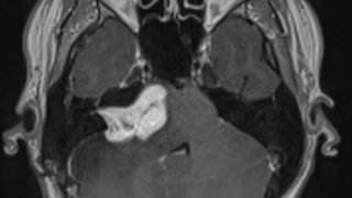
It is important to emphasize that “Moyamoya” is a Japanese word meaning “puff of smoke,” and it refers to how the new blood vessels that form look on an X-ray. Today, with advanced imaging such as CT and MRI, the vessels are much more defined and, while the “puff of smoke” is no longer seen, narrowed or occluded intracranial carotid arteries accompanied by small vascular networks are frequently referred to as “moyamoya findings.” They look the same, but the cause and patients’ ages are usually different.

MMD: The first signs of moyamoya disease usually appear as either a stroke or as recurrent transient ischemic attacks (TIAs) – when blood flow to the brain is altered temporarily – without the presence of other health conditions that might explain the cerebrovascular event.
Stroke and TIA can cause symptoms such as:
- Sudden, intense headache
- Vision changes
- Balance problems
- Numbness or weakness, often on one side of the body
- Trouble with focus or memory
TIAs end after a few minutes or hours but are considered a medical emergency. Sometimes called “mini or warning strokes,” TIAs can signal a serious problem with the brain circulation. If you experience symptoms of a stroke or TIA, call 911 immediately.
Females are twice as likely as males to develop MMD. Research is ongoing as to what causes MMD, which may derive from a mutation of the RNF213 gene that is involved in blood vessel development, according to the U.S. National Library of Medicine.
MMS: Stroke and TIA are also signs of MMS, which is the result of other health problems that lead to a similar picture of the blood vessel. These health problems include:
- Atherosclerosis (hardening of the arteries)
- Diabetes
- Down syndrome
- Hyperthyroidism
- Neurofibromatosis type 1
- Sickle cell anemia
- Smoking
When an adult 30 or older has a stroke, MMS and lifestyle risk factors are much more likely to be the cause than MMD, which affects only about 5 in 100,000 people who are predominantly of Asian descent.
Related reading: What to expect during stroke recovery
When stroke symptoms appear, BE FAST
Dr. Babu Welch talks about stroke, aneurysms, and the BE FAST formula that can help anyone recognize the symptoms of a stroke. UT Southwestern is 1 of 10 Advanced Comprehensive Stroke Centers in Texas and provides the highest standard of care for stroke victims.
Diagnosing and treating patients with moyamoya vessels
While MMD is a pediatric diagnosis, MMS sometimes can be tricky to diagnose. Vessel changes look similar to other aging-related diseases such as atherosclerosis, in which the arteries narrow due to a buildup of cholesterol and plaque. Getting an accurate diagnosis requires a team approach:
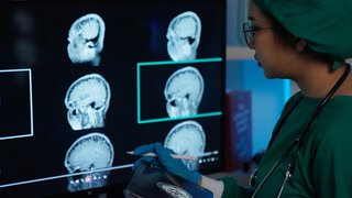
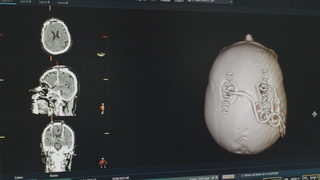
- Imaging: We start with computed tomography or magnetic resonance imaging (MRI) scans to look for the characteristic arterial narrowing and abnormal blood vessels, as well as any evidence of past strokes. Each of these types of studies can give us insight into the perfusion, or blood distribution, in the brain. Blood flow, volume, and transit time are affected by the narrowed blood vessels that are the result of moyamoya changes. It takes longer for blood to get to the right place when the blood vessels are small.
- Arteriography: If those studies suggest MMD or MMS, we then recommend a cerebral angiography, which creates images of the blood flow through the brain, to confirm the diagnosis.
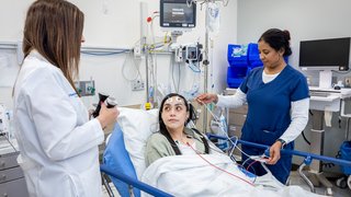
- Neuropsychology: We will test your memory and cognition (ability to think) to determine how your brain is being affected by too little blood. These tests serve as a baseline for post-treatment recovery.

Depending on the severity of your condition, your care team may recommend medical or surgical treatment. Neither MMD nor MMS can be cured, but treatment can reduce symptoms and prevent strokes. Every patient requires a personalized plan to achieve the safest, most effective treatment.
Medical management: Blood thinners (aspirin), calcium channel blockers, and anti-seizure drugs may be able to manage symptoms for a time. However, these medications won’t stop the blood vessels from narrowing. Lifestyle modifications that address diet and weight changes are very important in management. In patients where symptoms are frequent, surgery may also become necessary to ensure adequate blood flow to the brain.
Surgery: Moyamoya brain surgeries are delicate procedures that require an experienced cerebrovascular neurosurgeon. Two effective procedures include:
- Direct bypass, a type of revascularization in which we reroute blood flow around the narrowed artery by connecting the superficial temporal artery and middle cerebral artery – much like building a frontage road to the brain.
- Indirect bypass, which involves dissecting several inches of the superficial temporal artery and laying it onto the brain, where the tissue will grow new blood vessels to feed the brain over weeks to months. Frequently, the covering of the brain (dura) or muscle from the scalp can be used as another indirect blood donor. We use this approach only when direct bypass is not feasible. For example, in children whose arteries are not developed enough or are too small to withstand rerouting.
The expected outcome from bypass surgery at UT Southwestern is excellent. Most patients will have more than 97 percent unobstructed blood flow after the procedure, with the potential to dramatically reduce stroke risk.
Advances in brain surgery mean we no longer have to shave a patient’s entire head – just a strip where we can access the skull. Your vascular neurosurgeon will hide the incision in your hairline as best they can. After either surgery, you will need to stay in the hospital two to three days. Many patients can return to normal activities after four to six weeks.
Living with moyamoya disease or syndrome
If you have been diagnosed with MMD or if you have a condition associated with MMS, it’s important to know your personal risk factors and how to identify the signs of a stroke.
Most importantly, you must take care of your new source of blood to the brain. And that means identifying, controlling, and potentially preventing other health conditions that cause injury to or narrowing of your blood vessels.
Some patients with MMD or MMS will need to take blood thinners for life after surgery to reduce the risk of stroke. Talk with your doctor about the safest long-term stroke prevention options for you.
UT Southwestern endocrinologists, cardiologists, neurologists, and hematologists will work with you to optimize your medications, nutrition, and exercise habits for a heart-healthy life. To visit with a moyamoya disease or syndrome expert, call 214-645-8300 or request an appointment online.