How plasma exchange helped save her heart, decades after strep attacked her brain
February 18, 2026
New Patient Appointment or 214-645-7700
UT Southwestern Medical Center has earned a "High Performing" rating from U.S. News & World Report for aortic valve surgery, placing us among the nation’s top hospitals for this procedure.
UT Southwestern Medical Center provides expert care for patients with all types of heart valve disorders, from the common to the most complex. Our cardiac specialists use advanced diagnostic tools and leading surgical and interventional techniques to restore proper valve function, improve heart performance, and enhance quality of life.
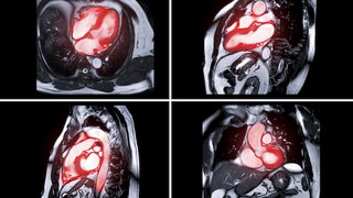
Four valves ensure blood flows in the right direction through the heart by opening and closing at the right time. When there’s a problem with a valve, there's a heart valve disorder.
Heart valve disorders make the heart work harder. As a result, patients may feel tired or short of breath. UT Southwestern’s heart experts are focused on fixing heart valve disorders so patients feel better and have more energy.
Valve disease treatment has improved dramatically over the past 15 years, from being able to replace faulty valves with prosthetics to surgically repairing the valves without cracking the chest (as is done in traditional open surgery).
The main valve conditions that disrupt the flow of blood in your heart are:
A third type of heart valve disorder, atresia, is when the valve is closed or doesn’t exist. It’s normally diagnosed within a few days of birth and requires immediate treatment.
Valve disorders can affect any of the four valves, and the name of the disorder reflects the valve that is causing the problem. The most common valve disorders are:
As you get older, your risk of getting a heart valve disorder increases. You may also be at higher risk if you’ve had a heart attack or heart failure or are at risk for coronary artery disease.
Some people with a heart valve disorder experience no symptoms. Those who do may have:
If your doctor hears a heart murmur by listening to your heart with a stethoscope, a valve problem might be the cause. The next step is a referral to a cardiologist for further examination and additional testing, which may include:
Heart valve disorders are treated either surgically or percutaneously (via a needle puncture through the skin). The goal is to repair or replace the valve, and this can be done in one of three ways: open surgery, minimally invasive surgery, or percutaneously. At UT Southwestern, we are experts in whichever treatment is appropriate for each patient, and we offer several innovative surgical options that patients won’t find anywhere else in North Texas.
Transcatheter aortic valve replacement (TAVR) is a minimally invasive procedure for patients with severe symptomatic aortic stenosis. After an evaluation by our heart team, we’ll recommend the best treatment (surgical or transcatheter) for each patient.
The TAVR procedure is performed by inserting a catheter through the groin to make a repair to the aortic valve. It is a far less invasive alternative to open-heart surgery. Mitrocep – Abbott’s MitraClip is a product used for this procedure.
Patients with mitral regurgitation and who are too old or sick for open surgery may be treated with the MitraClip. Going through the skin (percutaneously), we clip together the two leaflets of the mitral valve to stop the leaking.